Neuraxial Anesthesia in Patients with Scoliosis
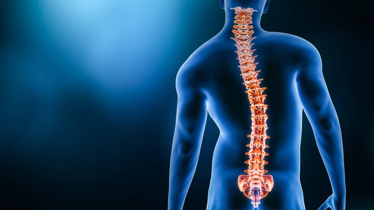
Scoliosis is an abnormal curvature of the spine in the coronal, axial, and/or sagittal planes. The condition is most frequently idiopathic and diagnosed in adolescence, but it can also be a congenital or neuromuscular condition. According to the National Scoliosis Foundation, scoliosis patients make up over 600,000 of private physician office visits, 30,000 children are fitted with a brace, and 38,000 patients undergo spinal fusion surgery per year.1 Naturally, this deformation of the spine can have an effect on the administration and spread of neuraxial, or spinal, anesthesia, with disparities in spread being a well-recognized complication for patients with scoliosis.
As reported by a pilot study by Ballarapu et al. in 2020 in the Indian Journal of Anaesthesia, many anesthesiologists are reluctant to administer neuraxial anesthesia in patients with scoliosis out of fear of postoperative complications (including potential neurological deficits), needing multiple attempts, and the unpredictability in the level and pattern of blockade during spinal anesthesia in these patients.2 Given the prevalence of scoliosis in our population and these known potential complications, it is important for surgeons and anesthesiologists to be familiar with the differences of neuraxial anesthesia in patients with scoliosis, as well as to work to develop a more sophisticated standard of care for these patients.
A 2013 paper in Regional Anesthesia by researchers from the Vanderbilt University School of Medicine reports on a case in which computerized tomography was used to assist in the placement of an epidural catheter in a patient with severe scoliosis and congenital dwarfism. An anesthesiologist typically administers neuraxial anesthesia by using spinal landmarks as guidance or by using ultrasound guidance. Due to the risks posed in trying to administer neuraxial anesthesia to a patient with such pronounced scoliosis (including neural injury, spinal hematoma, post-dural puncture headache, or infection as well as, more generally, a decrease in procedure efficiency and increase in patient discomfort and decrease in patient satisfaction), the computerized tomography data, corroborated by fluoroscopic images and ultrasound, were determined to be necessary precautions prior to operating on this patient. The surgical procedure was conducted without complications, and there were no adverse effects of the surgery. The authors suggested an algorithm to guide neuraxial techniques in scoliotic patients, taking the etiology and severity of a patient’s scoliosis into account in determining what techniques may be necessary in reducing risk of injury or discomfort for the patient in administering neuraxial anesthesia.3
A later (2016) report by Sharma and McConachie in Journal of Obstetric Anaesthesia and Critical Care reviewed further findings and techniques on how to safely administer neuraxial anesthesia in scoliotic patients. Noting that neuraxial blocks, despite being the preferred form of anesthesia for parturient patients in particular, were historically avoided in patients who had scoliosis or previous back surgery due to its comparative difficulty, higher chance of complications, and decreased efficacy, these authors also spotlight the use of ultrasound technology as a potential key element in overcoming these difficulties in providing neuraxial blocks to patients with scoliosis. They found through their review of recent literature that the use of ultrasound reduced the number of attempts and necessary levels of epidural catheter placement for all parturient patients, including those with a history of severe scoliosis or scoliosis repair. By allowing for the identification of interspinous spaces, ultrasound assisted with the insertion angle of the Tuohy needle and with the identification of the epidural space in these cases.4
All in all, as detailed in a 2020 review on the topic,5 ultrasound-guided placement of neuraxial blocks has become common practice for patients with severe scoliosis. Using this technique, it is possible to minimize the risk of misplacement, complications, or postoperative pain. Due to this technique becoming commonplace, and due to the prevalence of patients with either scoliosis or other conditions that increase the difficulty of administering neuraxial anesthesia (such as obesity, prior history of back surgery, or non-scoliotic abnormalities of the spine), it is important that anesthesiologists who routinely perform lumbar neuraxial blocks be familiar with both the sonoanatomy of the lumbar vertebrae and the techniques for ultrasound-guided placement of neuraxial blocks.
References
(1) Scoliosis – Symptoms, Diagnosis and Treatment. https://www.aans.org/en/Patients/Neurosurgical-Conditions-and-Treatments/Scoliosis.
(2) Ballarapu, G.; Nallam, S.; Samantaray, A.; Kumar, V. K.; Reddy, A. Thoracolumbar Curve and Cobb Angle in Determining Spread of Spinal Anesthesia in Scoliosis. An Observational Prospective Pilot Study. Indian J Anaesth 2020, 64 (7), 594. https://doi.org/10.4103/ija.IJA_914_19.
(3) Bowens, C.; Dobie, K. H.; Devin, C. J.; Corey, J. M. An Approach to Neuraxial Anaesthesia for the Severely Scoliotic Spine. Br J Anaesth 2013, 111 (5), 807–811. https://doi.org/10.1093/bja/aet161.
(4) Sharma, M.; McConachie, I. Neuraxial Blocks in Parturients with Scoliosis and after Spinal Surgery. J Obstet Anaesth Crit Care 2016, 6 (2), 70. https://doi.org/10.4103/2249-4472.191594.
(5) Yoo, S.; Kim, Y.; Park, S.-K.; Ji, S.-H.; Kim, J.-T. Ultrasonography for Lumbar Neuraxial Block. Anesth Pain Med 2020, 15 (4), 397–408. https://doi.org/10.17085/apm.20065.