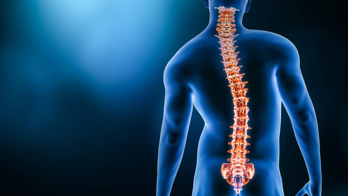
Calcium is crucial for various physiological processes, especially muscle contraction. In skeletal muscle, calcium release from the sarcoplasmic reticulum initiates an action potential leading to muscle contraction. Calcium also triggers contractions in smooth and cardiac muscle. Intercalated discs enable the transmission of contraction signals between cardiac myocytes, leading to synchronized muscle contractions throughout the heart. This coordination ensures that electrical impulses travel from the atria to the ventricles, driving blood flow through the circulatory system. Disruptions in calcium levels, such as hypocalcemia, can interfere with these normal physiological processes and impact anesthesia protocols.
Acute hypocalcemia (below 8.5 mg/dL) can cause syncope, congestive heart failure, numbness and tingling, bronchospasm and wheezing, laryngospasm and dysphagia, irritability, depression, fatigue, and seizures. The hallmark of acute hypocalcemia is tetany, characterized by neuromuscular irritability. Electromyographically, tetany involves repetitive, high-frequency discharges following a single stimulus. Peripheral neuron hyperexcitability is a significant pathophysiological effect of hypocalcemia, occurring at all levels of the nervous system, including motor endplates, spinal reflexes, and the central nervous system. Chronic hypocalcemia can result in coarse hair, brittle nails, psoriasis, dry skin, pruritus, poor dentition, and cataracts. Common physical exam findings of hypocalcemia include neural hyperexcitability, psychological disturbances, and cardiac arrhythmias.
The major factors influencing serum calcium concentration are parathyroid hormone (PTH), vitamin D, fibroblast growth factor 23 (FGF23), the calcium ion itself, and phosphate. Low serum calcium levels are most often caused by disorders of PTH or vitamin D. Hypocalcemia with low PTH occurs when there is decreased secretion of PTH due to destruction of the parathyroid glands (e.g. postsurgical, autoimmune), abnormal parathyroid gland development, or altered regulation of PTH production and secretion. Among the various causes of hypocalcemia, postsurgical hypoparathyroidism and autoimmune hypoparathyroidism are two of the more common ones. Hypocalcemia with high PTH occurs when PTH rises in response to low serum calcium levels to mobilize calcium from the kidneys and bones and to increase vitamin D production. Chronic hypocalcemia occurs when these actions are inadequate to restore serum calcium to normal levels. A high serum PTH in a patient with hypocalcemia may be secondary to vitamin D deficiency, chronic kidney disease, or pseudohypoparathyroidism, which is a rare condition.
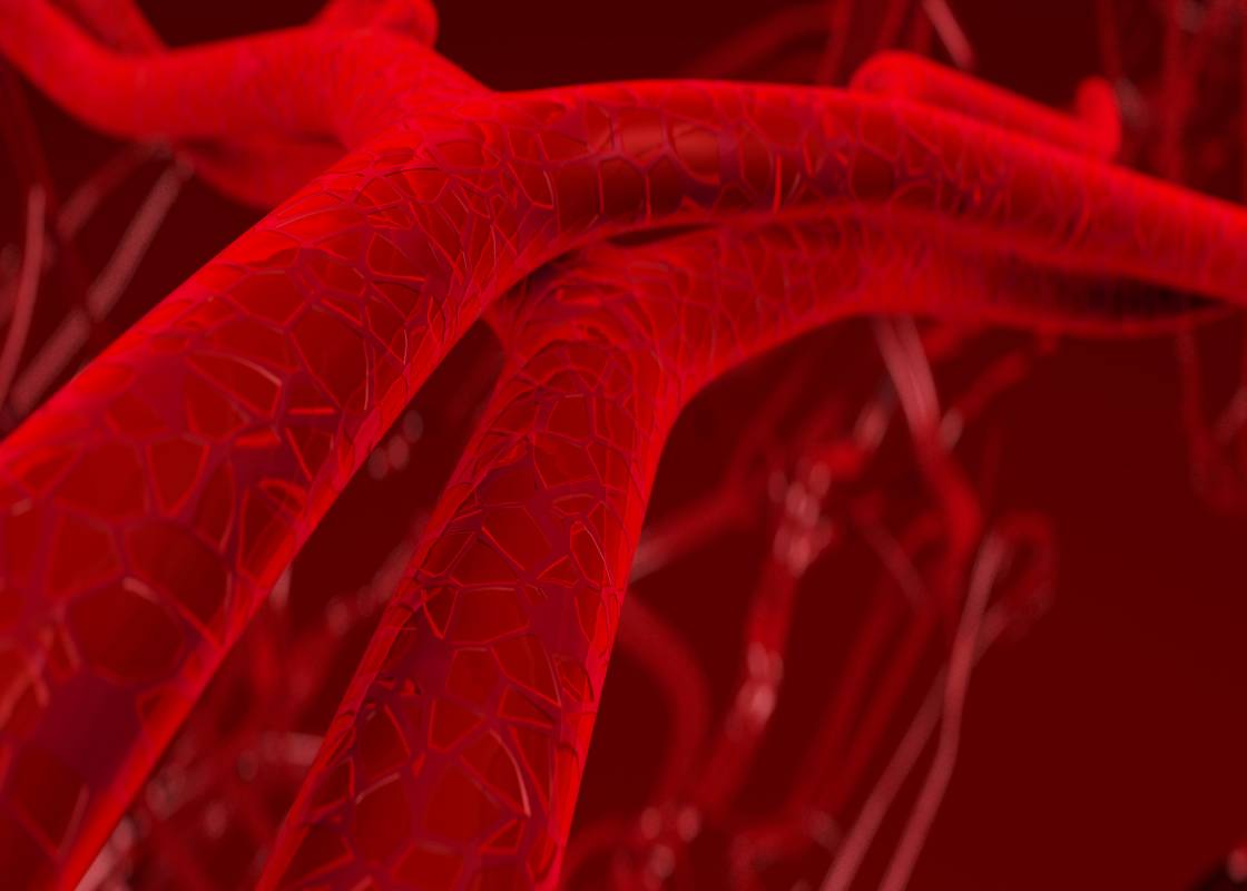
During anesthesia, various factors can alter serum ionized calcium levels, thereby increasing the risk of adverse effects from hypocalcemia in susceptible patients. These factors include malnutrition and low albumin levels, abnormal acid-base balance and electrolyte levels, medications used during the peri-operative period, transfusion of large volumes of citrated blood, and the use of cardiopulmonary bypass. Anesthetists should aim to prevent further changes in plasma calcium concentration and promptly recognize and treat the adverse effects of hypocalcemia, especially those affecting the heart. Preoperative evaluations typically include an assessment of electrolytes, including calcium, though abnormalities may potentially also arise intraoperatively or postoperatively.
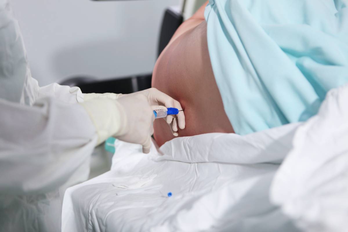
The treatment of hypocalcemia depends on its severity and underlying cause. Patients with severe symptoms (such as bronchospasm, seizures, or decreased cardiac function) require rapid correction with intravenous calcium therapy. For those with milder symptoms of neuromuscular irritability, initial treatment with oral calcium supplementation is sufficient. Intravenous calcium is also indicated to prevent acute hypocalcemia in patients with mild hypocalcemia or chronic hypocalcemia who cannot take or absorb oral supplements, often due to complex surgical procedures requiring prolonged recovery.
In conclusion, understanding the role of calcium in physiological processes, the diverse factors influencing its concentration, and the appropriate management strategies for hypocalcemia is essential for ensuring optimal patient care, particularly for anesthesia and surgery.
References
Riccardi D, Brown EM. Physiology and pathophysiology of the calcium-sensing receptor in the kidney. Am J Physiol Renal Physiol. 2010 Mar;298(3):F485-99. doi: 10.1152/ajprenal.00608.2009. PMID: 19923405; PMCID: PMC2838589.
Hannan FM, Thakker RV. Investigating hypocalcaemia. BMJ. 2013 May 9;346:f2213. doi: 10.1136/bmj.f2213. PMID: 23661111.
Desai TK, Carlson RW, Geheb MA. Prevalence and clinical implications of hypocalcemia in acutely ill patients in a medical intensive care setting. Am J Med. 1988 Feb;84(2):209-14. doi: 10.1016/0002-9343(88)90415-9. PMID: 3407650.
Zivin JR, Gooley T, Zager RA, Ryan MJ. Hypocalcemia: a pervasive metabolic abnormality in the critically ill. Am J Kidney Dis. 2001 Apr;37(4):689-98. doi: 10.1016/s0272-6386(01)80116-5. PMID: 11273867.
Kluger MT, Tham EJ, Coleman NA, Runciman WB, Bullock MF. Inadequate pre-operative evaluation and preparation: a review of 197 reports from the Australian incident monitoring study. Anaesthesia. 2000 Dec;55(12):1173-8. doi: 10.1046/j.1365-2044.2000.01725.x. PMID: 11121926.
Blitz JD, Kendale SM, Jain SK, Cuff GE, Kim JT, Rosenberg AD. Preoperative Evaluation Clinic Visit Is Associated with Decreased Risk of In-hospital Postoperative Mortality. Anesthesiology. 2016 Aug;125(2):280-94. doi: 10.1097/ALN.0000000000001193. PMID: 27433746.