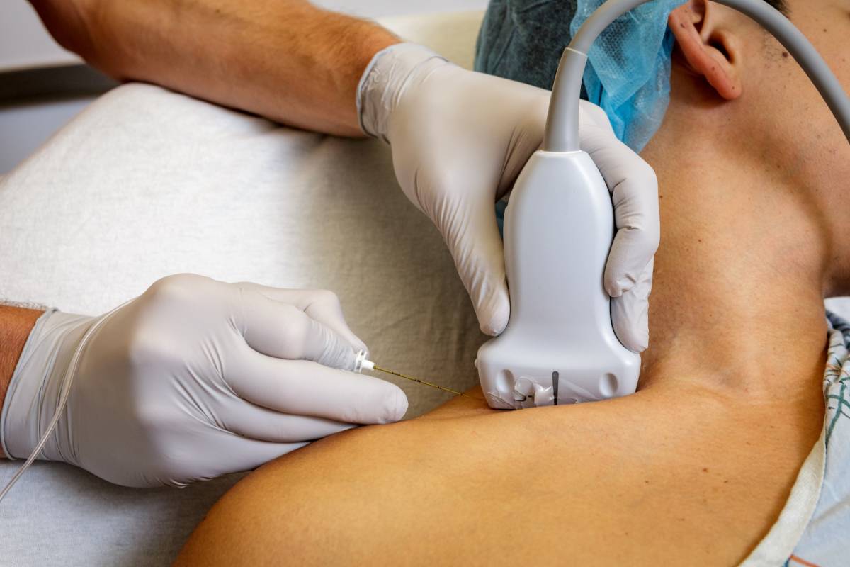
Anesthesia for Prostate Surgery

Prostate surgery can be carried out to treat prostate cancer, an enlarged prostate or benign prostatic hyperplasia (BPH), urinary continence, or erectile dysfunction in some cases 1. The goal of anesthesia for prostate surgery is to minimize pain before, during, and after surgery.  

There are multiple different approaches for prostate surgery, depending on the goal, which will require different anesthesia approaches. In a traditional open surgery or open approach, a surgeon will make an incision through the skin to excise out the prostate and nearby tissues. Either a radical retropubic or a radical perineal approach can be used during open prostate surgery. For both approaches, a patient can be under general anesthesia or spinal or epidural anesthesia.

Regional anesthesia and analgesia may favor minimal residual disease following removal of a primary tumor due to how it affects the body. A retrospective review of patient medical records looked at records from patients receiving open prostatectomy with general anesthesia. This study identified that, after adjusting for tumor size and date of surgery, among other factors,  using epidural analgesia with general anesthesia instead of postoperative opioids with general anesthesia was linked to a substantially lower risk of biochemical cancer recurrence after prostate surgery 2.  

A recent retrospective study revealed that combined spinal epidural anesthesia seems to be suitable and efficient for patients undergoing open radical retropubic prostatectomy. This specific approach reduces the average time in the operating unit and the length of the hospital stay 3

Laparoscopic surgery is a minimally invasive prostate surgery method that has become more widespread due to advancing technology and techniques. Two main approaches can be used tfor laparascopic prostate surgery. A laparoscopic radical prostatectomy requires multiple small cuts for the insertion of small surgical instruments, after which a surgical camera is inserted. In a robotic-assisted laparoscopic radical prostatectomy, a surgeon directs a robotic arm while viewing a computer monitor, providing maneuverability and precision. 

While any induction agent can be used for anesthesia, anesthetic management of prostate surgery patients needs to consider medical co-morbidities and medically optimize patients prior to surgery. Research has found that side effects remain minimal with robotic-assisted laparoscopic prostatectomy: Anesthetic and perioperative complications are rare. However, these do include a 1.3% incidence of postoperative anemia, postoperative pulmonary emboli, and, seemingly most commonly, corneal abrasions 4.  

Meanwhile, prostate laser surgery, endoscopic surgery, transurethral resection of the prostate (TURP), or transurethral incision of the prostate are additional surgical approaches that can help with urine flow. While TURP remains the surgical gold standard to treat benign prostatic hyperplasia, perioperative-associated morbidity remains high, ranging from 18% and 26%. In addition, between 1% and 8% of TURP procedures are complicated by TURP syndrome 5. The symptoms of TURP syndrome and bladder perforation may be masked under sedation and general anesthesia 6, and regional anesthesia may be warranted 7

Research has found that open prostatectomy may incur fewer complications if the prostate is very large (>100 g); prostatic size may be preemptively assessed by transrectal ultrasound scanning, endoscopic inspection, or manual examination 8

In any case, it is important after the surgery to keep the surgical wound clean, not drive, not do any high-energy activity, avoid sitting for more than 45 minutes, and take pain medications as prescribed. 

Additional research is required in order to validate and elucidate the links between anesthesia for prostate surgery and certain clinical outcomes. For example, prospective randomized trials to assess the link between substituting epidural analgesia for postoperative opioids and the substantially lower risk of cancer recurrence are required. This research will continue to help select the best anesthesia approach depending on clinical context. 

 
References 

1. What You Need to Know About Prostate Surgery. Available at: https://www.healthline.com/health/prostate-surgery. (Accessed: 3rd February 2023) 

2. Biki, B. et al. Anesthetic technique for radical prostatectomy surgery affects cancer recurrence: A retrospective analysis. Anesthesiology (2008). doi:10.1097/ALN.0b013e31817f5b73 

3. Kofler, O. et al. Anesthesia for Open Radical Retropubic Prostatectomy: A Comparison between Combined Spinal Epidural Anesthesia and Combined General Epidural Anesthesia. Prostate Cancer (2019). doi:10.1155/2019/4921620 

4. Danic, M. J. et al. Anesthesia considerations for robotic-assisted laparoscopic prostatectomy: A review of 1,500 cases. in Journal of Robotic Surgery (2007). doi:10.1007/s11701-007-0024-z 

5. Hahn, R. G. Fluid absorption in endoscopic surgery. British Journal of Anaesthesia (2006). doi:10.1093/bja/aei279 

6. Demirel, I., Ozer, A. B., Bayar, M. K. & Erhan, O. L. TURP syndrome and severe hyponatremia under general anaesthesia. BMJ Case Rep. (2012). doi:10.1136/bcr-2012-006899 

7. Mebust, W. K. et al. Transurethral prostatectomy: Immediate and postoperative complications. Cooperative study of 13 participating institutions evaluating 3,885 patients. J. Urol. (2002). doi:10.1016/S0022-5347(05)65370-0 

8. O’Donnell, A. M. & Foo, I. T. H. Anaesthesia for transurethral resection of the prostate. Contin. Educ. Anaesthesia, Crit. Care Pain (2009). doi:10.1093/bjaceaccp/mkp012 

Marburg Virus: What to Know

Recently identified as belonging to the filovirus genus, the Marburg virus (MARV) is another aggressive infectious agent with similar epidemiology to the Ebola virus (EBOV). There have been sporadic cases of MARV in Africa over the last decade, but the most recent cases occurred in Ghana this summer. Although this was Ghana’s first time encountering cases of Marburg virus, the quick response of medical practitioners at the clinic in the Ashanti region where the first case was identified was a key step in interrupting the spread of MARV throughout the region.2  

Ghana’s supposed patient zero was a 26-year-old male who eventually succumbed to the disease 3 days after his symptoms started on June 24, 2022. His symptoms included fever, malaise, epistaxis, bleeding from the mouth, and subconjunctival hemorrhage. On June 28, 2022, a 51-year-old male reported to the hospital with the same symptoms; he died the same day. Blood samples from both victims were tested using reverse transcriptase-polymerase chain reaction (RT-PCR) at the Noguchi Memorial Institute for Medical Research (NMIMR). NMIMR confirmed MARV as the cause of death. Further investigation by the Pasteur Institute in Senegal verified the results. According to the investigation conducted, animal contact played no role in these two cases. The evidence supports human to human transmission; the primary route is direct exposure to the blood or bodily fluids of an infected person. Contact tracing was used to identify 90 individuals who were exposed; they were quarantined and monitored.1 In total, 198 people were tested for MARV and all presented with negative results.3 Dr. Kasalo, W.H.O. representative to Ghana, credits the success of quelling this outbreak to “early alert and response, strong surveillance, community involvement and participation, and coordinated efforts”.2 W.H.O. declared an end of the Marburg virus outbreak September 16, 2022. 2  

Historically, MARV outbreaks on the continent have been moderate, regularly reporting less than 400 cases per incident. In 1980, Kenya reported 2 cases of MARV with fever and malaise. Seven years later, one additional case was reported in Kenya. From 1998-2000, the Democratic Republic of Congo dealt with 154 cases, but the worst outbreak happened in Angola between 2004-2005. A total of 374 cases were reported in this outbreak. Finally, Uganda had the most recent MARV cases and these periodically emerged from 2007-2017. No more than 15 cases were reported per outbreak in Uganda.1 

The CDC states that preventative measures against Marburg virus are poorly defined since no vaccine or antiviral drugs exist. Avoiding fruit bats, the natural host of the virus is recommended to prevent zootrophic transmission and early identification and isolation of a human host also prevents transmission. People at highest risk for contracting MARV are veterinarians working with non-human primates, laboratory workers handling the virus and travelers visiting regions of Africa endemic to fruit bats. To prevent infection in such settings, wearing personal protective clothing like gloves, masks, and gowns is recommended. Avoiding fecal matter from the host bats and infected humans is necessary, especially in the clinical setting. Rapid testing is recommended by the CDC to control the spread. 4 

References 

1.  Jack Wellington, Ayça Nur, Aderinto Nicholas, Olivier Uwishema, Hassan Chaito, Olutola Awosiku, Yusuf Jaafer Al Tarawneh, Jana Abdul Nasser Sharafeddine, Chinyere Vivian Patrick Onyeaka, Helen Onyeaka, “Marburg virus outbreak in Ghana: An impending crisis,” Annals of Medicine and Surgery, Volume 81, 2022,104377,ISSN 2049-0801 

2. Sayibu Ibrahim Suhuyini “Beating Marburg virus outbreak: Ghana’s journey to victory” https://www.afro.who.int/countries/ghana/news/beating-marburg-virus-outbreak-ghanas-journey-victory  

3. Mensah, Kent “Ghana Marburg Outbreak Declared Over” https://www.voanews.com/a/ghana-marburg-outbreak-declared-over-/6750533.html 

4. Center for Disease Control and Prevention “Marburg” https://www.cdc.gov/vhf/marburg/index.html  

Role of IV Fluids During Minor Surgery

Minor surgery is generally defined as a surgical procedure which does not require general anesthesia and can be performed electively and in an outpatient setting. Of note, there is no clear official delineation between major and minor surgeries, but minor surgeries are less invasive and less risky (Newsome et al., 2021). Patients undergoing any surgeries are often required to fast from fluid and food beforehand. This, however, can lead to dehydration and place them at risk of organ injury and failure. Administering intravenous (IV) fluids may have positive effects. Conversely, fluid overload may decrease pulmonary function and gut motility (Brandstrup, 2006). Most research on fluid therapy during surgery focuses on major surgery. However, less is understood on the value of IV fluids during minor surgery and how they may affect a patient’s clinical course.

A study found that patients who received 2 liters of IV fluids intra- and postoperatively recovered quicker from the effects of surgery and anesthesia compared to patients without fluids. This was a group of patients undergoing ambulatory gynecologic laparoscopy surgery. Though this is a small volume of IV fluids, it appeared to have immediately measurable effects on patients postoperatively. The authors hypothesized that this was due to a correction of  dehydration. In terms of patient satisfaction, 93% of patients who had intravenous fluids felt the most recent anesthetic experience was better than past ones (Keane & Murray, 2007).  Other trials testing different intravenous fluid volumes on the outcomes of outpatient surgery found similar improvements in self-reported symptoms such as drowsiness and dizziness. In general, the volume of fluids given was approximately the same as the deficit from fasting, which may point to a benefit to replacing fluid losses from fasting. However, this research did not show if IV fluids would be beneficial in minor surgery in terms of external loss of fluid during an operation (Brandstrup, 2006).  

However, patient satisfaction, though important, is a subjective assessment of IV fluids during minor surgery. In two groups of 15 patients undergoing minor gynecologic surgery, there was no obvious clinical benefit of IV fluids administration (Ooi et al., 1992). This study attempted to use objective testing including two tests of psychomotor function. There was no significant difference between postoperative motor reaction times. Notably, the patients in this study were healthy and underwent a short duration of anesthesia. Fluids and hydration may be more important for longer surgeries. 

Furthermore, the benefit of IV fluids during minor surgery may depend on a patient’s risk level. Preoperative administration of 2mL/kg for every hour patients had fasted from fluids decreased incidence and severity of postoperative nausea and vomiting. However, this was for patients scheduled for diagnostic gynecologic laparoscopy, which is typically a less urgent procedure compared to other situations that would require minor surgery (Maharaj et al., 2005). Other factors that may influence recovery include nature and duration of the procedure, individual patient risk, and the anesthesia method used (Ooi et al., 1992).  

Ooi et al. brought up an interesting point in their study: perhaps further research may focus on the benefits of oral fluids versus IV fluids in minor surgery. Oral fluids offer a more cost-effective solution, and patients are often deprived of fluids a long period before minor surgeries (Ooi et al., 1992). Clear fluids may be safe for patients in limited volumes closer to the surgery start time. Another topic of research may focus on the effects of fluid overload compared to hypovolemia. This topic may fuel further investigation into more carefully assessing role of fluids during minor surgery.  

References 

Brandstrup B. Fluid therapy for the surgical patient. Best Pract Res Clin Anaesthesiol. 2006;20(2):265-283. doi:10.1016/j.bpa.2005.10.007 

Keane PW, Murray PF. Intravenous fluids in minor surgery. Their effect on recovery from anaesthesia. Anaesthesia. 1986;41(6):635-637. doi:10.1111/j.1365-2044.1986.tb13059.x 

Maharaj CH, Kallam SR, Malik A, Hassett P, Grady D, Laffey JG. Preoperative intravenous fluid therapy decreases postoperative nausea and pain in high risk patients. Anesth Analg. 2005;100(3):675-682. doi:10.1213/01.ANE.0000148684.64286.36 

Newsome K, McKenny M, Elkbuli A. Major and minor surgery: Terms used for hundreds of years that have yet to be defined. Ann Med Surg (Lond). 2021;66:102409. Published 2021 May 25. doi:10.1016/j.amsu.2021.102409 

Ooi LG, Goldhill DR, Griffiths A, Smith C. IV fluids and minor gynaecological surgery: effect on recovery from anaesthesia. Br J Anaesth. 1992;68(6):576-579. doi:10.1093/bja/68.6.576

IV Lidocaine for Surgical Pain

Intravenous lidocaine is widely used for its effect on postoperative pain and recovery. In addition, concern about opioid risks in the postoperative period has galvanized the use of nonopioid analgesic adjuncts.1 However, if used inappropriately and incorrectly, intravenous lidocaine can have fatal consequences – therefore, it is extremely important to administer intravenous (IV) lidocaine for surgical pain according to a careful, well-informed protocol.

The decision to administer intravenous lidocaine or not depends on the type of surgery and individual patient factors, including but not limited to the presence of any existing condition that affects pain management and risk (such as pre-existing chronic pain). This decision mainly focuses on three priorities. First, is intravenous lidocaine safe? Second, does intravenous lidocaine effectively reduce postoperative pain and speed up recovery? Third, how is intravenous lidocaine licensed for use?2

In general, while perioperative IV lidocaine infusion is indeed effective at reducing pain, evidence supporting its use varies according to the surgical procedure. However, the benefits of intravenous lidocaine are clear in certain clinical contexts. For example, it prevents airway reactivity on emergence in smokers and quenches cerebral hemodynamic responses to airway manipulation.3 It may also reduce anesthetic requirements by approximately one-third in specific situations.4 It may further reduce neuropathic pain by inhibiting the activity of injured afferent nerves.5 Finally, following laparoscopic nephrectomy, it can reduce the need for postoperative morphine, ameliorating postoperative pain management and recovery.6

In general, notable guidelines have been developed to ensure the appropriateness, safety and efficacy of intravenous lidocaine for surgical pain.  

First and foremost, the use of IV lidocaine for acute surgical pain should be approved by the local hospital and medication governance committee or equivalent. When possible, consent should also be obtained by patients.  

As regards its administration, certain researchers recommend an initial dose not exceeding 1.5 mg/kg, calculated using the patient’s ideal body weight and provided as an infusion over 10 minutes. Thereafter, researchers recommend an infusion not exceeding 1.5 mg/kg/h for no more than 24 hours, subject to re-assessment.  

Furthermore, intravenous lidocaine should not be used in conjunction with any other local anesthetic interventions. Therefore, intravenous lidocaine should not be administered within 4 hours of any nerve block. Conversely, no nerve block should be performed within 4 hours of discontinuing an intravenous lidocaine infusion. 

Outside the operating theater and recovery room, patients receiving intravenous lidocaine should be monitored to quickly address complications, if any arise. Particular attention should be paid to patients who have an existing comorbidity.2

In the end, however, the approach selected for the use of intravenous lidocaine should be approved by hospital health governance systems, and the individual clinical decision should be carried out following properly informed consent on behalf of the patient. 

In conclusion, IV lidocaine may be a key pillar of a pain management strategy for surgical pain. However, it needs to be very carefully delivered. In the meantime, additional research in the form of further randomized control trials with a large sample size7 is warranted in order to corroborate and specify current protocols.

References 

1. Dunn, L. K. & Durieux, M. E. Perioperative Use of Intravenous Lidocaine. Anesthesiology (2017). doi:10.1097/ALN.0000000000001527 

2. Foo, I. et al. The use of intravenous lidocaine for postoperative pain and recovery: international consensus statement on efficacy and safety. Anaesthesia (2021). doi:10.1111/anae.15270 

3. Hamill, J. F., Bedford, R. F., Weaver, D. C. & Colohan, A. R. Lidocaine before endotracheal intubation: Intravenous or laryngotracheal? Anesthesiology (1981). doi:10.1097/00000542-198111000-00016 

4. Kaba, A. et al. Intravenous lidocaine infusion facilitates acute rehabilitation after laparoscopic colectomy. Anesthesiology (2007). doi:10.1097/00000542-200701000-00007 

5. Kirillova, I. et al. Effect of local and intravenous lidocaine on ongoing activity in injured afferent nerve fibers. Pain (2011). doi:10.1016/j.pain.2011.02.046 

6. Tauzin-Fin, P. et al. Benefits of intravenous lidocaine on post-operative pain and acute rehabilitation after laparoscopic nephrectomy. J. Anaesthesiol. Clin. Pharmacol. (2014). doi:10.4103/0970-9185.137269 

7. Yue, H., Zhou, M., Lu, Y., Chen, L. & Cui, W. Effect of intravenous lidocaine on postoperative pain in patients undergoing intraspinal tumor resection: Study protocol for a prospective randomized controlled trial. J. Pain Res. (2020). doi:10.2147/JPR.S249359

Prone Position for Surgery

Surgery in the prone position, or lying face down, as opposed to supine where the patient is lying on their back with their face up, is used when a surgery requires access to anatomical structures posteriorly. This can include access to the posterior head, neck, or spine during a spinal surgery or even for access to the upper urinary tracts during urological surgeries [1].

Prone surgery is associated with many complications that often stem from the prolonged and increased pressure on anterior organs. Increased abdominal pressure causes excess pressure to build on the inferior vena cava, the major vein which returns blood from the body back to the heart. This results in a back-up of blood in the body and thus, less blood returning to the heart. Further, the pressure on the chest wall leads to decreased output from the heart which results in a subsequent drop in blood pressure. This drop in blood pressure can be dangerous in the setting of surgery. Additionally, respiratory rate is affected in prone surgery, which can lead to further decreases in cardiac output and oxygenation [2]. Some of the potential cardiovascular complications of prone surgery include hypovolemia and cardiac arrest [1]. Increased bleeding is also a common complication of the prone position, especially in spinal surgery, as damage to engorged veins can cause greater bleeding than damage to normal veins [2].

Prone positioning can also increase intraocular pressure [3]. Malpositioning of the patient can lead to direct pressure on the eye for an extended period, which can lead to irreversible visual loss. While postoperative vision loss is commonly described in journals, the true rate of incidence remains low [1]. However, while rare, it is necessary for both surgeons and anesthesiologists to watch for adequate patient eye protection during prone surgeries to avoid this potentially devastating complication.  

There are many preoperative risk factors that play a role in postoperative complications of prone surgery. In terms of postoperative vision loss, older patients who have an elevated intraocular pressure at baseline typically are at higher risk for vision loss [3]. Hypertension, diabetes, obesity, anemia, atherosclerosis, and history of smoking are all risk factors for postoperative complications [5].

Ideally in a prone surgery, positioning will maximize ventilation of the patient while minimizing risk of bleeding and damage to vital organs [2]. Over the years, special operating tables and other devices such as chest rolls and bolsters have been designed to promote ideal prone positioning, decrease pressure on the abdomen and chest, and lower complications associated with prone positioning. Further, careful positioning can prevent vision loss by changing the degree of the bed so that the head is above the heart. This allows for less venous pooling of blood in the eye and orbit which can help to decrease intraocular pressures [4]. Reducing the length of time patients are in prone positioning can also help to decrease complications. Surgeries that are longer than 6.5 hours are considered prolonged, and it may be beneficial to use a series of shorter surgeries to reduce risks of complications. On the other hand, risks of multiple surgeries may outweigh the risks of one prolonged prone surgery [3,5]. Overall, for preventing complications of prone surgery, it is important to monitor fluids, using IV fluids when necessary to replace intravascular volume, monitor blood loss and hemoglobin level, and optimize blood pressure within 20-25% of baseline blood pressure [5,6].  

References 

  1. Kwee, Melissa M., et al. “The Prone Position during Surgery and Its Complications: A Systematic Review and Evidence-Based Guidelines.” International Surgery, vol. 100, no. 2, Feb. 2015, pp. 292–303, www.ncbi.nlm.nih.gov/pmc/articles/PMC4337445/, 10.9738/intsurg-d-13-00256.1. 
  1. Schonauer, Claudio, et al. “Positioning on Surgical Table.” European Spine Journal, vol. 13, no. S01, 22 June 2004, pp. S50–S55, 10.1007/s00586-004-0728-y. 
  1. ‌van Wicklin, Sharon Ann. “Systematic Review and Meta-Analysis of Prone Position on Intraocular Pressure in Adults Undergoing Surgery.” International Journal of Spine Surgery, 14 Apr. 2020, p. 7029, 10.14444/7029. 
  1. Ozcan, Mehmet S., et al. “The Effect of Body Inclination during Prone Positioning on Intraocular Pressure in Awake Volunteers: A Comparison of Two Operating Tables.” Anesthesia & Analgesia, vol. 99, no. 4, Oct. 2004, pp. 1152–1158, 10.1213/01.ane.0000130851.37039.50.  
  1. ‌Shifa, Jemal, et al. “A Case of Bilateral Visual Loss after Spinal Cord Surgery.” Pan African Medical Journal, vol. 23, 2016, 10.11604/pamj.2016.23.119.8443.  
  1. Lee, Lorri A., et al. “The American Society of Anesthesiologists Postoperative Visual Loss Registry.” Anesthesiology, vol. 105, no. 4, 1 Oct. 2006, pp. 652–659, 10.1097/00000542-200610000-00007.  

Implications of the Telehealth Modernization Act

The Telehealth Modernization Act was passed in 2022, modifying requirements for the coverage of telehealth services under Medicare. It effectively extends certain flexibilities initially authorized during the COVID-19 public health emergency. Endorsed by a number of professional organizations, from the American Association of Orthopedic Surgeons (AAOS) (1) to the American Hospital Association (AHA) (2), the legislation has a number of critical implications.

In April 2020, at the beginning of the pandemic, over 40% of Medicare fee-for-service primary care visits were carried out through telehealth, and over 10 million beneficiaries accessed telehealth services from mid-March through early July of 2020 (3). While regulatory coverage restrictions have long prevented telehealth services for many of the nation’s roughly 61 million Medicare beneficiaries, the COVID-19 pandemic acutely highlighted the importance of access to telehealth care.

Among other elements, the bill allows (1) federally qualified health centers and rural health clinics to act as the location of the health care practitioner; (2) the home of a beneficiary to act as the location of the health care beneficiary for all services; and (3) all types of practitioners to provide telehealth services, as assessed by the Centers for Medicare and Medicaid Services (4).  

The Telehealth Modernization Act applies to a full spectrum of health care. In addition to providing Medicare recipients many additional telehealth services, the act will 1) help patients continue to access telehealth from varied health care workers, from speech language pathologists to physical therapists, and 2) help Medicare patients receiving services ranging from home dialysis to hospice care keep receiving this care through telehealth. 

The most important implication of the Telehealth Modernization Act is the increased flexibility of – and therefore access to – health care. Permanently eradicating geographic and originating site restrictions, the Telehealth Modernization Act will reduce access barriers that have continuously increased in recent times. Relatedly, it will enable greater access to high-quality health care services for vulnerable populations, in particular among seniors.  

In addition to ensuring access to essential care for patients, the increased use of telehealth helps address certain workforce pressures, including those that arose as a result of an overwhelmed healthcare system during the COVID-19 pandemic.  

Many opportunities and challenges remain to be addressed in view of a telehealth-centric future, however (5). First, patient trust and security are top priorities (6). One study found that ensuring that health services provision meets patients’ needs at all times depends not only on a certain degree of flexibility in care delivery modalities and interprofessional collaboration, but also healthy, sustained relationships with patients. Second, and relatedly, it will be important to ensure that patients receive the right amount of empathic, personalized care, crucial to a good patient experience and patient well-being. As many have experienced, virtual interactions so far have major differences compared to in-person ones. To this end, guidelines and training are needed, as well as careful attention to technological challenges and interpersonal relationship needs (7).  

Overall, telehealth has been beneficial for millions of Americans, especially during the pandemic (3). While continued research is required to ensure that its key provisions are implemented in the most smooth and efficient way possible, the Telehealth Modernization Act will surely continue to positively reshape healthcare delivery.  

References 

1. AAOS’ Advocacy Efforts Focus on Access to Quality Care [Internet]. [cited 2022 Jul 25]. Available from: https://www.aaos.org/aaosnow/2022/may/advocacy/advocacy01/ 

2. AHA Comments to Modernization Subcommittee of the Healthy Future Task Force Re: Telehealth | AHA [Internet]. [cited 2022 Jul 25]. Available from: https://www.aha.org/lettercomment/2022-03-04-aha-comments-modernization-subcommittee-healthy-future-task-force-3-4-22 

3. Scott, Schatz, Shaheen Introduce Bipartisan Legislation to Increase Access to Telehealth in the Midst of the Pandemic | U.S. Senator Tim Scott of South Carolina [Internet]. [cited 2022 Jul 25]. Available from: https://www.scott.senate.gov/media-center/press-releases/scott-schatz-shaheen-introduce-bipartisan-legislation-to-increase-access-to-telehealth-in-the-midst-of-the-pandemic 

4. H.R.1332 – 117th Congress (2021-2022): Telehealth Modernization Act | Congress.gov | Library of Congress [Internet]. [cited 2022 Jul 25]. Available from: https://www.congress.gov/bill/117th-congress/house-bill/1332 

5. Blandford A, Wesson J, Amalberti R, AlHazme R, Allwihan R. Opportunities and challenges for telehealth within, and beyond, a pandemic. The Lancet Global Health. 2020. doi: 10.1016/S2214-109X(20)30362-4. 

6. Hale TM, Kvedar JC. Privacy and security concerns in telehealth. Virtual Mentor. 2014. doi: 10.1001/virtualmentor.2014.16.12.jdsc1-1412. 

7. Breton M, Sullivan EE, Deville-Stoetzel N, McKinstry D, DePuccio M, Sriharan A, et al. Telehealth challenges during COVID-19 as reported by primary healthcare physicians in Quebec and Massachusetts. BMC Fam Pract. 2021; doi: 10.1186/s12875-021-01543-4

Viral Shedding in Vaccinated Individuals

Viral shedding refers to the expulsion of live virus from an organism in which the virus has successfully replicated.1 Shedding can occur via several different mechanisms, such as budding, whereby viral particles exit the host cell membrane by surrounding themselves with molecules from the membrane, and apoptosis, whereby viral particles force cells to undergo cell death and can then be released into the extracellular space.2 The amount of viral shedding by infected individuals depends on the virus in question, the stage of infection, and the host’s immunity against the virus, including if they have been vaccinated.

 Vaccination can protect individuals from viral infection such that even upon exposure to a virus, the individual’s vaccine-induced immunity kills the infectious agent before it can replicate and cause illness. In reality, breakthrough infections can also occur, in which vaccinated individuals contract a viral illness. Vaccinated individuals who contract breakthrough infections may nevertheless have better outcomes than unvaccinated individuals who become infected. Variants of SARS-CoV-2, such as the Delta variant, have caused breakthrough infections, but it has been found that people vaccinated against COVID-19 are much less likely than unvaccinated individuals to develop severe symptoms.3 

Does this mean that fully vaccinated individuals are also less likely to transmit the virus to others – do they shed less virus? Dr. Jiwon Jung and other South Korean researchers recently published their findings on this very question. In a recent Journal of the American Medical Association paper, Jung et al. reported on their study of the transmissibility of COVID-19 according to vaccination status.4 The study involved two cohorts of vaccinated and unvaccinated individuals. The first cohort was subject to a secondary transmission study: the contacts of the cohort members were tracked, and their COVID-19 infections were recorded. The second cohort underwent a viral kinetics shedding study: patients submitted saliva samples each day of the study, and the viral load in the samples was measured using polymerase chain reaction (PCR). 

The results from both cohorts indicate that vaccinated individuals with breakthrough infections shed less virus than unvaccinated infected patients. The risk of secondary transmission was significantly lower in the breakthrough group than in the non-breakthrough group. Additionally, while patients in the breakthrough and non-breakthrough had a similar initial viral load after infection, fully vaccinated patients had a shorter duration of viral shedding compared to partially vaccinated and unvaccinated individuals. 

Though convincing, the study, as its authors acknowledge, has several limitations. Perhaps most significantly, viral shedding was only measured by the amount of virus in saliva samples. Due to “logistical challenges,” nasopharyngeal swab samples were not used, though they could have enabled more accurate data on viral shedding, especially given the incomplete data regarding the amount of viral shedding at different body sites over the course of infection.5 Furthermore, the study only tracked nosocomial secondary transmission – that is, infections originating in a hospital – which may bias the results. Finally, the cohort 1 study began before the emergence of the infectious Delta variant but concluded after it was widespread. The study did not control for SARS-CoV-2 variant, and the conclusions drawn from it are therefore limited. Nevertheless, this important study hopefully paves the way for future research into viral shedding in vaccinated individuals. 

References 

1. Yan, D. et al. Characteristics of Viral Shedding Time in SARS-CoV-2 Infections: A Systematic Review and Meta-Analysis. Front. Public Health 9, (2021), DOI: 10.3389/fpubh.2021.652842 

2. Badu, K. et al. SARS-CoV-2 Viral Shedding and Transmission Dynamics: Implications of WHO COVID-19 Discharge Guidelines. Front. Med. 8, (2021), DOI: 10.3389/fmed.2021.648660 

3. CDC. COVID-19 Vaccination. Centers for Disease Control and Prevention https://www.cdc.gov/coronavirus/2019-ncov/vaccines/effectiveness/why-measure-effectiveness/breakthrough-cases.html (2020)

4. Jung, J. et al. Transmission and Infectious SARS-CoV-2 Shedding Kinetics in Vaccinated and Unvaccinated Individuals. JAMA Netw. Open 5, e2213606 (2022), DOI:10.1001/jamanetworkopen.2022.13606 

5. Congrave-Wilson, Z. et al. Change in Saliva RT-PCR Sensitivity Over the Course of SARS-CoV-2 Infection. JAMA 326, 1065–1067 (2021), DOI: 10.1001/jama.2021.13967 

Epinephrine as an Adjuvant to Anesthetics

While nerve blocks are widely used alongside anesthesia, its effect may last only up to a few hours, and increasing the dose to prolong the block may result in adverse side effects on the cardiovascular or central nervous systems 1. Therefore, an adjuvant to anesthetics is often used because of its synergistic effect with anesthetics, prolonging the duration of sensory-motor blocks and limiting the dose requirement of local anesthetics. Over time, local anesthetics of choice have evolved from classical opioids to a number of drugs with varying mechanisms of action 2. Epinephrine, opioids, alpha-2 adrenergic antagonists, steroids, anti-inflammatory drugs, midazolam, and ketamine, among others, have been used to augment the effect of local anesthetics. Among these, epinephrine has been commonly used as an adjuvant to anesthetics for a few reasons. 

One of the oldest additives to local anesthetic solutions, epinephrine potentiates local anesthetics through its anti-nociceptive properties mediated by alpha-2 adrenoreceptor activation 3, alongside its vasoconstrictive properties which limit the systemic reabsorption of local anesthetics and thus prolong anesthetic action 4. It has a recommended dosing of 0.5-1.0 μg/kg 5

One study found that using lidocaine with high-dose epinephrine (200 μg/mL) for an axillary block prolonged motor block and sensory block by approximately 25-40 minutes but increased the incidence of tachycardia and hypertension. A lower dose of 25 μg/mL, however, had a minimal effect, prolonging a motor block by only 10 minutes and a sensory block by just 30 minutes 6. Another study found that, when added alongside mepivacaine for a brachial plexus block, epinephrine prolonged a motor and sensory block by approximately one hour 7. However, epinephrine has not been found to prolong blockade with ropivacaine and does not match up to other adjuvants, such as clonidine, in the prolongation of a brachial plexus block with bupivacaine 8,9.  

In addition, as an adjuvant, epinephrine can be added to local anesthetics to detect intravascular injection. As such, it has played a significant role in preventing the inadvertent intravascular administration of local anesthetic solutions. However, the recent surge in the routine use of ultrasonography in nerve blocks has decreased its utility in these contexts.  

A few limitations on the use of epinephrine as an adjuvant to anesthetics remain. Not only may perineural administration of epinephrine incur detrimental effects to the nerves of patients who are smokers or who suffer from diabetes mellitus or hypertension 10, but also epinephrine in too high a dose may be neurotoxic.  

A lower dose of epinephrine might be just as effective at prolonging its anesthetic effects while limiting the hazards associated with its unintentional intravascular injection. To this end, one study concluded that 1.25 μg/mL of epinephrine, when coinjected with clinical doses of bupivacaine and levobupivacaine, resulted in a comparable vasoconstrictor effect in human skin to that of higher concentrations 11.  

Further research is warranted to investigate how to address ongoing challenges linked to the use of epinephrine as an adjuvant to anesthetics. In this context, research will particularly need to focus on minimizing its deleterious effects.  

References 

1. Jeon, Y. H. The use of adjuvants to local anesthetics: Benefit and risk. Korean Journal of Pain (2018). DOI: 10.3344/kjp.2018.31.4.233

2. Swain, A., Nag, D. S., Sahu, S. & Samaddar, D. P. Adjuvants to local anesthetics: Current understanding and future trends. World J. Clin. Cases (2017). DOI: 10.12998/wjcc.v5.i8.307

3. Collins, J. G., Kitahata, L. M., Matsumoto, M., Homma, E. & Suzukawa, M. Spinally administered epinephrine suppresses noxiously evoked activity of WDR neurons in the dorsal horn of the spinal cord. Anesthesiology (1984). DOI: 10.1097/00000542-198404000-00001

4. Cazaubon, Y. et al. Population pharmacokinetics of articaine with 1:200,000 epinephrine during third molar surgery and simulation of high-dose regimens. Eur. J. Pharm. Sci. (2018). DOI: 10.1016/j.ejps.2017.11.027

5. Jöhr, M. & Berger, T. M. Caudal blocks. Paediatric Anaesthesia (2012). DOI: 10.1111/j.1460-9592.2011.03669.x

6. Dogru, K. et al. Hemodynamic and blockade effects of high/low epinephrine doses during axillary brachial plexus blockade with lidocaine 1.5%: A randomized, double-blinded study. Reg. Anesth. Pain Med. (2003). DOI: 10.1016/S1098-7339(03)00225-6

7. Song, J. H. et al. Comparison of dexmedetomidine and epinephrine as an adjuvant to 1% mepivacaine in brachial plexus block. Korean J. Anesthesiol. (2014). DOI: 10.4097/kjae.2014.66.4.283

8. Weber, A., Fournier, R., Van Gessel, E., Riand, N. & Gamulin, Z. Epinephrine does not prolong the analgesia of 20 mL ropivacaine 0.5% or 0.2% in a Femoral three-in-one block. Anesth. Analg. (2001). DOI: 10.1097/00000539-200111000-00060

9. Eledjam, J. J. et al. Brachial plexus block with bupivacaine: effects of added alpha-adrenergic agonists: comparison between clonidine and epinephrine. Can. J. Anaesth. (1991). DOI: 10.1007/BF03036962

10. Myers, R. R. & Heckman, H. M. Effects of local anesthesia on nerve blood flow: Studies using lidocaine with and without epinephrine. Anesthesiology (1989). DOI: 10.1097/00000542-198911000-00021

11. Newton, D. J., McLeod, G. A., Khan, F. & Belch, J. J. F. The effect of adjuvant epinephrine concentration on the vasoactivity of the local anesthetics bupivacaine and levobupivacaine in human skin. Reg. Anesth. Pain Med. (2004). DOI: 10.1016/j.rapm.2004.04.011

Medical License Regulations and Reciprocity

Medical licenses are a key regulatory measure which determines who can (and can’t) practice medicine. A medical license can only be obtained once certain basic requirements have been met, including a doctorate from an accredited U.S. or international medical school, a certificate of completion of residency, a passing score on the licensing examination, and demonstration of mental, moral, and physical fitness to practice medicine. Many licensing boards even limit the number of attempts that can be made to pass the licensing examinations, known as USMLE for medical doctorate candidates or COMLEX-USA for osteopathic doctorate candidates [1]. These steps alone take years to complete and thus regulations leave only a select few who qualify as candidates for a license.

Yet even once obtained, medical licenses are highly regulated when it comes to where and how they can be used. Medical licenses are administered by individual states, not the federal government. As such, the regulations around obtaining and maintaining a medical license is different throughout different U.S. states. Examples of key variances include the minimum number of years of postgraduate residency, or the number of times one is allowed to retake the licensing examination. Because of the variance in qualifications between states, doctors in the U.S. are often geographically limited to their primary state of practice. However, increased mobility of physicians and prevalence of telehealth services have led to a rise in doctor-patient relationships across state borders. Minute differences in license regulations can therefore pose a large obstacle when it comes to access to care.  

In light of this, some states have implemented medical license reciprocity, which allows for expedited licensure to practice in another state. Essentially, these states have agreed to coordinate the necessary requirements to receive and maintain a medical license, therefore enabling physicians who participate in the Interstate Medical Licensure Compact to more easily practice across state lines. As of August 2021, 32 states/territories were participating in medical license reciprocity [2], including: 

Alabama, Arizona, Colorado, Delaware, Georgia, Guam, Idaho, Illinois, Iowa, Kansas, Kentucky, Louisiana, Maine, Maryland, Michigan, Minnesota, Mississippi, Montana, Nebraska, Nevada, New Hampshire, North Dakota, South Dakota, Oklahoma, Texas, Tennessee, Utah, Vermont, West Virginia, Wisconsin, Wyoming, and Washington 

Several more have passed the IMLC but are currently in the process of implementation or are experiencing implementation delays.  

Just as medical licenses must be earned differently in different states, the grounds for license revocation varies by state. However, in this regard there are more similarities than differences. Doctors may lose their license if they demonstrate they no longer have the mental, physical, or moral capacity to practice safely. For example, some causes of license revocation in California are substance abuse, insurance fraud, patient abuse, violating drug prescription laws, loss of records/violation of HIPAA, and discrimination [3].

Given the surge in popularity of telehealth appointments resulting from the COVID-19 pandemic, patients have new expectations concerning the availability of their most trusted physicians, even if that means that the online appointment must take place across state lines. As a result, it is likely that more states will look to join the Interstate Medical Licensure Compact in the years to come. 

References 

1. About physician licensure. FSMB. (n.d.). Retrieved from https://www.fsmb.org/u.s.-medical-regulatory-trends-and-actions/guide-to-medical-regulation-in-the-united-states/about-physician-licensure/#:~:text=All%20state%20medical%20boards%20require,training%20to%20obtain%20a%20license.  

2. Compact State Map. Interstate Medical Licensure Compact. (2021, August 20). Retrieved from https://www.imlcc.org/participating-states/  

3. California, S. of. (2022.). Laws. Medical Board of California. Retrieved from https://www.mbc.ca.gov/About/Laws/California-Law/  

The Role of General Anesthetics in Post-Operative Cognitive Dysfunction (POCD)

Post-operative cognitive dysfunction (POCD) is an expansive definition for the wide spectrum of cognitive deficits seen in patients after anesthesia and surgery. Most common deficits of POCD include cognitive impairments, such as memory and information processing, which can also be present in the aftermath of anesthetics. Current data suggests that POCD is most directly caused by neuroinflammation and microglial activation, which triggers an immuno-hormonal cascade increasing the permeability of the blood-brain barrier, ultimately promoting the influx of macrophages into the brain parenchyma. The pro-inflammatory cytokines synthesized by these macrophages cause neuronal damage, apoptosis, and impaired neurotransmission to the hippocampus, medial prefrontal cortex, amygdala, and the cingulate cortex, brain regions critical in memory, reward, emotion, and judgement [1-3].  

Eckenhoff et. al.’s research in 2004 demonstrated clinically relevant concentrations of inhaled anesthetics such as halothane and isoflurane could induce the oligomerization of amyloid β (peptides that compose the amyloid plaques in Alzheimer’s patients) and amyloid-β-induced cytotoxicity [4]. However, intravenous anesthetics such as ethanol and propofol did not have this effect. While desflurane (another inhalational agent) alone does not induce caspase-3 activation or affect amyloid precursor protein cells, a combination of desflurane and hypoxia does both [5]. Additionally, abnormal tau tangles are one of the hallmarks of AD. In 2007, a murine study found anesthetics including chloral hydrate, pentobarbital and isoflurane produced rapid and massive tau protein hyperphosphorylation and inhibition of Ser/Thr phosphatase (PP) activity. These researchers confirmed PP2A (a subtype of PPs) is the main phosphatase driving tau dephosphorylation; their inhibition is a direct result of hypothermia induced by anesthesia [6,7]. Another preclinical study found the inhaled anesthetic sevoflurane generated behavioral deficits in spatial learning and memory in aged rats; the rats also experienced hippocampal alterations, most notably a downregulation of the cAMP/CREB signaling pathway, a pathway extensively implicated as a “hot spot” of long-term potentiation, memory, and neuroprotection [8].  

Following the use of intravenous agents, specific neurological and behavioral changes have been reported. A prolonged infusion of propofol significantly impaired spatial learning in mice, as measured by the Morris water maze behavioral assessment. The CA1 region of the hippocampus showed significant autophagy inhibition, leading to the observed cognitive deficits [9]. Autophagy is the fundamental catabolic process involving degradation of dysfunctional cellular molecules to supply energy and compounds for further biosynthesis. Defective autophagy is associated with increased aging as well as diseases like cancer or neurodegenerative and muscular disorders [10]. Inhaled anesthetics also affect this process, as sevoflurane may cause impaired memory performance and increase hippocampal neuronal apoptosis through its suppression of autophagic processes, potentially increasing the risk of POCD [11]. However, rapamycin, an immunosuppressive drug, reduced the cognitive effects of sevoflurane by inducing autophagy [9,11].

POCD in human patients is often thought to be induced by increased activity of GABAA receptors, which most general anesthetics act on. The subtypes of GABAA receptors may be responsible for the different effects of general anesthesia, such as amnesia, sedation, and hypnosis [13]. Preclinical investigations have pinpointed the alpha-5 subunit of GABAA receptors to be especially responsible, given their significant upregulation after administration of GABAergic anesthetics [12]. Sevoflurane may damage synaptic plasticity by decreasing postsynaptic density protein in mPFC, highlighting another potential mechanism ultimately generating POCD [14].

Although research has proposed plausible mechanisms by which general anesthetics may lead to POCD, interesting perspectives for future consideration involve in-depth study of predisposing and precipitating factors of POCD.

References 

  1. Wang, B., Li, S., Cao, X., Dou, X., Li, J., Wang, L., Wang, M., & Bi, Y. (2017). Blood-brain barrier disruption leads to postoperative cognitive dysfunction. Current Neurovascular Research, 14(4), 359–367. https://doi.org/10.2174/1567202614666171009105825 
  1. Vacas, S., Degos, V., Feng, X., & Maze, M. (2013). The neuroinflammatory response of postoperative cognitive decline. British Medical Bulletin, 106, 161–178. https://doi.org/10.1093/bmb/ldt006  
  1. Cascella, M., & Bimonte, S. (2017). The role of general anesthetics and the mechanisms of hippocampal and extra-hippocampal dysfunctions in the genesis of postoperative cognitive dysfunction. Neural Regeneration Research, 12(11), 1780–1785. https://doi.org/10.4103/1673-5374.219032  
  1. Eckenhoff, R. G., Johansson, J. S., Wei, H., Carnini, A., Kang, B., Wei, W., Pidikiti, R., Keller, J. M., & Eckenhoff, M. F. (2004). Inhaled anesthetic enhancement of amyloid-β oligomerization and cytotoxicity. Anesthesiology, 101(3), 703–709. https://doi.org/10.1097/00000542-200409000-00019  
  1. Zhang, B., Dong, Y., Zhang, G., Moir, R. D., Xia, W., Yue, Y., Tian, M., Culley, D. J., Crosby, G., Tanzi, R. E., & Xie, Z. (2008). The inhalation anesthetic desflurane induces caspase activation and increases amyloid β-protein levels under hypoxic conditions *. Journal of Biological Chemistry, 283(18), 11866–11875. https://doi.org/10.1074/jbc.M800199200  
  1. Planel, E., Richter, K. E. G., Nolan, C. E., Finley, J. E., Liu, L., Wen, Y., Krishnamurthy, P., Herman, M., Wang, L., Schachter, J. B., Nelson, R. B., Lau, L.-F., & Duff, K. E. (2007). Anesthesia leads to tau hyperphosphorylation through inhibition of phosphatase activity by hypothermia. Journal of Neuroscience, 27(12), 3090–3097. https://doi.org/10.1523/JNEUROSCI.4854-06.2007  
  1. Nicolson, S. C., Montenegro, L. M., Cohen, M. S., O’Neill, D., Calfin, D., Jones, L. A., & Jobes, D. R. (2010). A comparison of the efficacy and safety of chloral hydrate versus inhaled anesthesia for sedating infants and toddlers for transthoracic echocardiograms. Journal of the American Society of Echocardiography: Official Publication of the American Society of Echocardiography, 23(1), 38–42. https://doi.org/10.1016/j.echo.2009.11.019  
  1. Xiong, W.-X., Zhou, G.-X., Wang, B., Xue, Z.-G., Wang, L., Sun, H.-C., & Ge, S.-J. (2013). Impaired spatial learning and memory after sevoflurane–nitrous oxide anesthesia in aged rats is associated with down-regulated cAMP/CREB signaling. PLOS ONE, 8(11), e79408. https://doi.org/10.1371/journal.pone.0079408  
  1. Yang, N., Li, L., Li, Z., Ni, C., Cao, Y., Liu, T., Tian, M., Chui, D., & Guo, X. (2017). Protective effect of dapsone on cognitive impairment induced by propofol involves hippocampal autophagy. Neuroscience Letters, 649, 85–92. https://doi.org/10.1016/j.neulet.2017.04.019  
  1. Papáčková, Z., & Cahová, M. (2014). Important role of autophagy in regulation of metabolic processes in health, disease and aging. Physiological Research, 63(4), 409–420. https://doi.org/10.33549/physiolres.932684  
  1. Zhang, X., Zhou, Y., Xu, M., & Chen, G. (2016). Autophagy is involved in the sevoflurane anesthesia-induced cognitive dysfunction of aged rats. PLOS ONE, 11(4), e0153505. https://doi.org/10.1371/journal.pone.0153505  
  1. Zurek, A. A., Yu, J., Wang, D.-S., Haffey, S. C., Bridgwater, E. M., Penna, A., Lecker, I., Lei, G., Chang, T., Salter, E. W. R., & Orser, B. A. (2014). Sustained increase in α5GABAA receptor function impairs memory after anesthesia. The Journal of Clinical Investigation, 124(12), 5437–5441. https://doi.org/10.1172/JCI76669  
  1. Bonin, R. P., & Orser, B. A. (2008). GABAA receptor subtypes underlying general anesthesia. Pharmacology Biochemistry and Behavior, 90(1), 105–112. https://doi.org/10.1016/j.pbb.2007.12.011 
  1. Ling, Y., Ma, W., Yu, L., Zhang, Y., & Liang, Q. (2015). Decreased PSD95 expression in medial prefrontal cortex (mPFC) was associated with cognitive impairment induced by sevoflurane anesthesia. Journal of Zhejiang University-SCIENCE B, 16(9), 763–771. https://doi.org/10.1631/jzus.B1500006