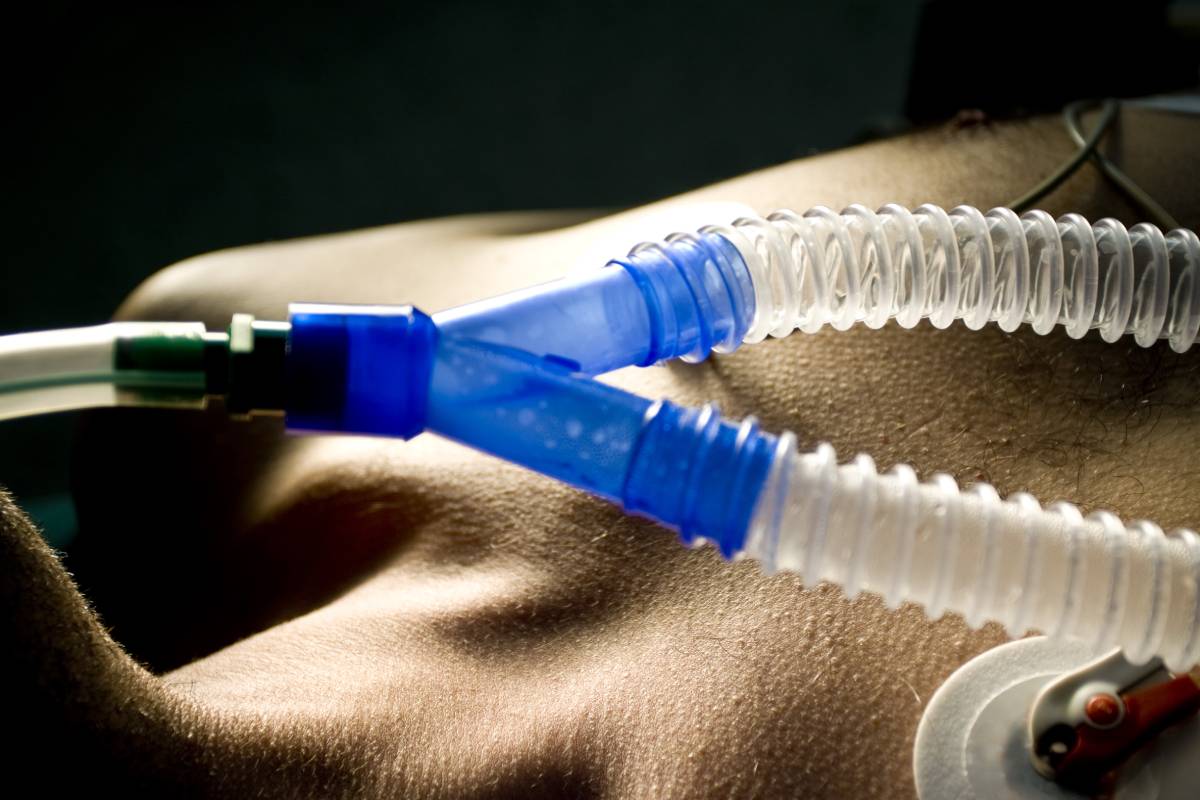
Indications for Nasotracheal Intubation

Nasotracheal intubation (NTI) is a common form of airway management [1]. During NTI, an endotracheal tube enters the patient’s trachea, following the pathway of the narrow nasal cavity [1]. Physicians need to be well aware of the indications and contradictions of NTI to ensure a favorable outcome for the patient [1]. According to those indications, special precautions or an alternate form of airway management may be necessary [1].

Multiple types of surgeries call for the use of nasotracheal intubation [1]. Often, NTI is used on patients undergoing oral and maxillofacial surgery, given the improved surgical field and mobility that it offers physicians during such operations [1]. More specifically, NTI can be appropriate for intranasal, oropharyngeal, mandible, dental, orthognathic, and rigid laryngoscope and orthognathic surgery [1, 2]. It can also be the airway management procedure of choice for patients receiving tonsillectomies, as well as for those undergoing “complex intra-oral procedures” that require mandibular reconstruction [2, 3].

Beyond certain types of surgery, other situations also benefit from the use of NTI. For one, patients who, due to trismus, have a limited mouth opening, should avoid orotracheal intubation and, therefore, turn to nasotracheal intubation [1]. Additionally, patients who suffer from either cervical spine degeneration or instability may benefit from NTI [1].

Anesthesia providers must also consider a variety of contraindications before deciding on nasotracheal intubation as their strategy of choice. Patients with basilar skull fractures, either with or without corresponding cerebrospinal fluid leakage, could experience severe trauma in their frontal lobes during NTI [1]. Coagulopathy can also place patients at risk of complication via severe epistaxis [1]. Other contraindications for NTI include midface instability, prosthetic heart valves, frequent epistaxis, epiglottitis, and nasal bone fracture [2]. If a patient has foreign bodies in their nasal passage, such as sizeable polyps, or has undergone recent nasal surgery, they may also not be a suitable candidate for NTI [2]. More recently, Zhu et al. found evidence to suggest that having a retropharyngeal internal carotid artery can narrow the pharyngeal cavity and even increase the risk of a tear during intubation [3]. Consequently, it may be beneficial for anesthesia providers to consider RICA before intubation to determine whether the nasotracheal technique is appropriate.

Technical and tube-related contraindications should also be considered. In terms of tube factors, physicians should carefully prevent the selection of too big of a tube, heightened cuff pressure, and the co-existing presence of a nasogastric tube [4]. As for technology, poor larynx visualization, several intubation attempts, and forceful intubation may also compromise the success of nasotracheal intubation [4].

To successfully identify indications and contraindications before deciding on NTI as the correct course of action, physicians should first engage in a meticulous pre-anesthesia evaluation within 48 hours of surgery [4]. During that time, it may become clear which side of the nose should be the one through which the endotracheal tube is passed [4]. However, if the physician remains undecided following the evaluation, a physical exam or an anterior rhinoscopy can be helpful [4]. To reduce the risk of complications, physicians can use a softened or smaller tube, as well as adrenaline to constrict the patient’s blood vessels, depending on the patient’s unique risk factors [5].

In the end, developing a patient-specific strategy that takes into account all relevant indications and contraindications, and applies techniques to meet those needs, is essential to successful nasotracheal intubation.

References

[1] D. H. Park et al., “Nasotracheal intubation for airway management during anesthesia,” Anesthesia and Pain Medicine, vol. 16, no. 3, p. 232-247, July 2021. [Online]. Available: https://doi.org/10.17085/apm.21040.

[2] T. B. Folino, G. McKean, and L. J. Parks et al., “Nasotracheal Intubation,” Treasure Island (FL): StatPearls Publishing, Updated January 19, 2021. [Online]. Available: https://www.ncbi.nlm.nih.gov/books/NBK499967/.

[3] W-P. Zhu et al., “Retropharyngeal internal carotid artery: a potential risk factor during nasotracheal intubation,” Surgical and Radiologic Anatomy, p. 1-8, June 2021. [Online]. Available: https://doi.org/10.1007/s00276-021-02784-9.

[4] D. Prasanna and S. Bhat, “Nasotracheal Intubation: An Overview,” Journal of Maxillofacial Oral Surgery, vol. 13, no. 4, p. 366-372, October-December 2014. [Online]. Available: https://doi.org/10.1007/s12663-013-0516-5.

[5] D. G. Canpolat and S. O. Yasli, “Does a Nasal Airway Facilitate Nasotracheal Intubation or Not? A Prospective, Randomized, and Controlled Study,” Journal of Oral and Maxillofacial Surgery, vol. 79, no. 1, p. 89.e.1-89.e.9, January 2021. [Online]. Available: https://doi.org/10.1016/j.joms.2020.08.029.

Health Incentives: Lessons for COVID-19 Vaccine Campaigns

While most countries still lack adequate access to COVID-19 vaccines, in the U.S., vaccine supply has begun to outstrip demand. As of July 2021, nearly 70 percent of adults have received at least one shot.1 But vaccination rates vary significantly across the country, and in some regions, opposition to vaccines is entrenched, with estimates of “hesitancy” and “strong hesitancy” hovering at one-third of a state’s population.2 To promote public health efforts — and to draw customers back through their doors — businesses have begun to offer incentives for vaccinated customers: Krispy Kreme made headlines in March 2021 when it announced free donuts for anyone who could prove they had been inoculated, and the New York Yankees and Mets are offering free tickets to fans who get vaccinated at ballparks before games. Local governments are approving six-digit budgets for vaccine perks and giveaways, and many states are instituting incentives-based programs as well: Maryland is paying fully vaccinated state employees $100, and in West Virginia, 16- to 35-year-olds who get vaccinated can receive $100 savings bonds.3

In spite of the popular appeal of vaccine incentives, some scholars have questioned their ethics. In a JAMA Network paper published in July 2021, Govind Persad and Dr. Ezekiel Emanuel respond to worries about coercion and exploitation by arguing that vaccination’s benefits — among them preventing harm from COVID-19 and protecting disadvantaged populations facing barriers to vaccination — do not threaten to deprive anyone of anything to which they were entitled. They do validate the legitimacy of two particular concerns: the potential to waste public funds and the potential to make vaccination look riskier than it is by offering compensation. They write that the former could be addressed by carefully calibrating benefits to what is needed to encourage vaccination and that the latter could be addressed by targeting vaccine incentives to only the most receptive groups.4

However, designing incentive programs that strike this balance is notoriously difficult. Positive incentives have proven to be effective in other health campaigns. For example, participants in a fitness-based employee wellness program who received a mere $20 monthly incentive maintained higher levels of exercise than non-participants over a 3-year period, after adjusting for pre-intervention differences in activity levels. However, those who had previously exercised the least were also the least likely to join the program.5 The results of studies like this one and others6,7 indicate that rewards-based incentives can be powerful tools for swaying certain segments of the population to change their behaviors, but that those most entrenched in their practices or beliefs may not be reached so easily.

An Axios-Harris poll from May 2021 found that nearly one-third of unvaccinated Americans say they’ll either “get the vaccine whenever they get around to it” or “will wait awhile and see before getting the vaccine” — suggesting that with well-strategized incentivization, the U.S. can continue to narrow the gap toward heard immunity.3 However, the question stands as to how to persuade the most staunchly opposed groups.

The backlash prompted by Israel’s brief consideration of a vaccination mandate is indicative of why tactical public health education programs are necessary.8 Though Israel acquired a sufficient supply of vaccines early on, many groups’ distrust of the vaccine compelled the state to implement a “Green Pass,” in which proof of vaccination or recovery from COVID-19 serves as a passport to communal events and spaces.9 The program’s punitive edge — barring unvaccinated individuals from the social, commercial, and cultural opportunities they may crave — comes with its own controversies. For example, Mexico’s tax on sugar-sweetened beverages has been criticized as an unfair instance of a government overstepping its place. On the other hand, Mexico’s sugar tax reduced purchases by nearly 10% in 2015.10 The tax is projected to prevent 239,900 cases of obesity and 61,340 cases of diabetes, which a Health Affairs article estimates saves $3.98 per dollar spent on the tax’s implementation.11 As for Israel’s Green Pass, over 80 percent of the adult population has now been fully vaccinated.9 Incentives can be powerful, but they must be implemented alongside robust public-health education campaigns.

References 

  1. The New York Times. See how vaccinations are going in your county and state. The New York times. https://www.nytimes.com/interactive/2020/us/covid-19-vaccine-doses.html. Published July 16, 2021. 
  1. Vaccine hesitancy for COVID-19: State, county, and local estimates. U.S. Department of Health and Human Services. Published June 17, 2021. https://aspe.hhs.gov/pdf-report/vaccine-hesitancy 
  1. Ducharme J. From free beer to $100 payments, states are incentivizing COVID-19 vaccination. Will it work? Time. Published online May 5, 2021. https://time.com/6046238/covid-19-vaccine-incentives/ 
  1. Persad G, Emanuel EJ. Ethical considerations of offering benefits to COVID-19 vaccine recipients. JAMA. Published online 2021. doi:10.1001/jama.2021.11045
  1. Crespin DJ, Abraham JM, Rothman AJ. The effect of participation in an incentive-based wellness program on self-reported exercise. Prev Med. 2016;82:92-98. 
  1. Strohacker K, Galarraga O, Williams DM. The impact of incentives on exercise behavior: a systematic review of randomized controlled trials. Ann Behav Med. 2014;48(1):92-99. 
  1. Burns RJ, Donovan AS, Ackermann RT, Finch EA, Rothman AJ, Jeffery RW. A theoretically grounded systematic review of material incentives for weight loss: implications for interventions. Ann Behav Med. 2012;44(3):375-388. 
  1. Wilf-Miron R, Myers V, Saban M. Incentivizing vaccination uptake: The “Green Pass” proposal in Israel. JAMA. 2021;325(15):1503-1504. 
  1. Kershner I. With most adults vaccinated and case numbers low, Israel removes many restrictions. The New York Times. Published June 1, 2021. https://www.nytimes.com/2021/06/01/world/middleeast/israel-covid-restrictions.html.
  1. Colchero MA, Rivera-Dommarco J, Popkin BM, Ng SW. In Mexico, evidence of sustained consumer response two years after implementing A sugar-sweetened beverage tax. Health Aff (Millwood). 2017;36(3):564-571. 
  1. Basto-Abreu A, Barrientos-Gutiérrez T, Vidaña-Pérez D, et al. Cost-effectiveness of the sugar-sweetened beverage excise tax in Mexico. Health Aff (Millwood). 2019;38(11):1824-1831. 

Ophthalmic Surgery Advancements and Changes in Anesthesia Strategies

Ophthalmic surgery is one of the most common surgical fields requiring anesthesia in developed countries; in the United States alone, over 3 million cataract lenses are extracted annually 1. In the future, ophthalmic surgeries will only continue to increase in frequency given the high incidence of age-related ophthalmic ailments in an overall aging population 2,3.

Either topical, regional or general anesthesia can be used for eye surgery, depending on patient background and clinical context. Topical anesthesia usually involves the administration of a topical anesthetic such as xylocaine, paracaine, tetracaine, or bupivacaine 4; however, it remains limited by its tendency to trigger allergies, endothelial and epithelial toxicity, or keratopathy. Local anesthesia is usually preferable for ophthalmic surgery since it is economical, safe, easy, has a rapid onset, and yields a dilated pupil with low intraocular pressure, in addition to incurring minimal post-operative restlessness, lung complications, and bleeding. Regional anesthesia can be used for nearly all eye surgeries, such as keratoplasty, cataract extraction, glaucoma, iridectomy, strabismus, and retinal detachment surgeries. For regional anesthesia as such, the conjunctiva, globe and orbicularis are paralyzed by a combination of surface and facial anesthesia, alongside, traditionally, a retrobulbar block. Rarely used, general anesthesia is preferred for ocular surgeries in anxious individuals, psychiatric patients, and youth – as well as for major surgeries such as exenteration or perforating ocular injuries. In such contexts, general anesthesia is advantageous in that it results in total akinesia, controlled intraocular pressure, and a safe operating environment.

In the past decade, as a result of drastic technological improvements, the practice of ophthalmic anesthesia has been transformed. In general, vast improvements have been made to minimally invasive surgery, primarily for glaucoma and cataracts 5–7, by refining smaller incisions for surgery. These improvements have laid the foundation for multiple changes to surgical and anesthesia approaches for opthalmic surgery.

First, resorting to general anesthesia is increasingly infrequent. Second, phacoemulsification techniques have gained prominence in light of their ability to facilitate the conduction of cataract surgery under topical anesthesia and to avoid the venous air embolisms that can be caused by suture-free vitrectomy ports; their advent has resulted in topical anesthesia representing a viable alternative for most cataract surgeries. Third, the sub-Tenon’s blockhas gained popularity given its ability to produce satisfactory anesthesia for most intraocular procedures and its circumvention of the inherent risks of needle-based blocks, including globe perforation and optic nerve injury. It is primarily used for cataract surgery but remains effective in a variety of other eye surgeries, including strabismus correction, vitreoretinal surgery, and trabeculectomy 8. Finally, the retrobulbar block has been largely replaced by the peribulbar approach 9, whereby needles are kept at a greater distance from vital adnexa and globe. Since peribulbar anesthesia has a prolonged latency of action, it is preferable to perform the block at least ten minutes prior to the start of surgery; these are increasingly performed in the holding room to increase efficiency.

In parallel, certain surgical techniques have also emerged as useful additions to these surgical approaches. For example, ultrasound-guided eye blocks have proven useful during ophthalmic surgeries. The potential benefits of ultrasound guidance include the real-time visualization of needle trajectory, thereby minimizing the chance of globe perforation 9, and the tracking of anesthetic spread, thereby improving the quality and safety of blocks. In the same vein, hyaluronidase, despite its allergenicity, has emerged as a useful adjuvant during surgery for its promotion of local anesthetic diffusion and hastening of block onset time. Teaching curricula will have to be updated to ensure the complete training of anesthesiologists as the field evolves 9. Of critical importance in light of an aging population, anesthesia strategies in ophthalmic surgery ostensibly continue to rapidly progress and dynamically adapt to ongoing technological advancements.

References

1.        Cullen KA, Hall MJ, Golosinskiy A. Ambulatory surgery in the United States, 2006. Natl Health Stat Report. 2009.

2.        Colby SL, Ortman JM. Projections of the Size and Composition of the U.S. Population: 2014 to 2060. Program. 2014.

3.        Swenor BK, Ehrlich JR. Ageing and vision loss: looking to the future. Lancet Glob Heal. 2021. doi:10.1016/S2214-109X(21)00031-0

4. Local Anaesthesia for Eye Surgery. https://web.archive.org/web/20121025132106/http://www.nda.ox.ac.uk/wfsa/html/u12/u1205_01.htm.

5.        Yang S-A, Mitchell W, Hall N, et al. Trends and Usage Patterns of Minimally Invasive Glaucoma Surgery in the United States. Ophthalmol Glaucoma. 2021. doi:10.1016/j.ogla.2021.03.012

6.        Singh K, Misbah A, Saluja P, Singh A. Review of manual small-incision cataract surgery. Indian J Ophthalmol. 2017. doi:10.4103/ijo.IJO_863_17

7.        De Gregorio A, Pedrotti E, Russo L, Morselli S. Minimally invasive combined glaucoma and cataract surgery: clinical results of the smallest ab interno gel stent. Int Ophthalmol. 2018. doi:10.1007/s10792-017-0571-x

8.        Guise P. Sub-Tenon’s anesthesia: An update. Local Reg Anesth. 2012. doi:10.2147/LRA.S16314

9.        Palte HD. Ophthalmic regional blocks: Management, challenges, and solutions. Local Reg Anesth. 2015. doi:10.2147/LRA.S64806

Estimated Disease Burden from Long Work Hours

In May of 2021, the World Health Organization (WHO) and International Labour Organization (ILO) presented the first joint estimate of the burden of work-related disease and injury. The report, published in Environment International, draws on data from 194 countries from 2000 to 2016 and finds that there are higher risks of ischemic heart disease and stroke among people who work long hours (55 hours per week or more) when compared to those who work 35-40 hours per week.

WHO/ILO estimates that over 745,000 deaths and 23.3 million disability-adjusted life years (DALYs) are attributable to ischemic heart disease and stroke. These findings are calculated using models called population attributable fractions (PAFs), which quantify the proportion of death or disease that is driven by a specific risk factor, such as overwork, based on extensive systematic reviews. Stroke was found to have a higher PAF than heart disease, with 6.9 percent of stroke deaths and 9.3 percent of stroke DALYs attributable to overwork, compared to 3.7 percent of heart disease deaths and 5.3 percent of heart disease DALYs.1

The disease burdens attributed to overwork are greater than those attributed to many other occupational hazards. The Global Burden of Disease (GBD) 2016 study found that 1.53 million deaths were attributable to a total of seven different risk factors (carcinogens; asthmagens; particulate matter, gases and fumes; secondhand smoke; noise; ergonomic risk factors for low back pain; and risk factors for injury). Notably, the study reported an overall reduction of deaths and DALYs since 1990, indicating that occupational exposures are controllable and that their proper regulation can prevent disease and death.2

But while exposure to some occupational hazards may be decreasing, between 2010 and 2016, the proportion of the population working long hours increased substantially — by 9.3 percent — according to the WHO/ILO report. This means that, based on the study’s models, deaths from ischemic heart disease and stroke increased by 41.5 percent and 19.0 percent, respectively. Those most affected include older men: nearly three-quarters of deaths occurred among males, and most of the deaths were recorded among people aged 60-79 years who had worked for 55 hours or more per week between the ages of 45 and 74 years. People living in the Western Pacific and Southeast Asia regions were also among those most likely to work long hours;3 in the U.S., less than 5 percent of the population works long hours.1

Notably, there has been some criticism of the WHO/ILO’s methodology, with authors of similar studies noting that the results are not supported by other published data reviews, and that subgroup analyses reveal greater complexity in patterns. In particular, Kivimäki et al. note that “findings of socioeconomic interaction suggest that at this stage the conclusion should be restricted to low socioeconomic status occupations only,” in response to a paper published in 2020 analyzing the effect of long working hours on ischemic heart disease.4 That is, the authors suggest that it is not only the length of working hours that impact health, but also the nature of the work being conducted. The WHO/ILO report concedes that more research must be done on a global scale to assess this hypothesis.1

The health consequences created by an overworked population is a subject that deserves increased scrutiny moving forward. Other scholars have suggested that economic recessions such as the 2008 global financial crisis can drive increases in working hours, as might the expansion of the gig economy and new working-time arrangements that include remote work. The WHO/ILO report only includes data from before the COVID-19 pandemic, and more research analyzing how this public health crisis continues to impact working conditions is warranted.

References 

  1. Pega F, Náfrádi B, Momen NC, et al. Global, regional, and national burdens of ischemic heart disease and stroke attributable to exposure to long working hours for 194 countries, 2000–2016: A systematic analysis from the WHO/ILO Joint Estimates of the Work-related Burden of Disease and Injury. Environ Int. 2021;(106595):106595. 
  1. GBD 2016 Occupational Risk Factors Collaborators. Global and regional burden of disease and injury in 2016 arising from occupational exposures: a systematic analysis for the Global Burden of Disease Study 2016. Occup Environ Med. 2020;77(3):133-141. 
  1. Long working hours increasing deaths from heart disease and stroke: WHO, ILO. World Health Organization. https://www.who.int/news/item/17-05-2021-long-working-hours-increasing-deaths-from-heart-disease-and-stroke-who-ilo 
  1. Kivimäki M, Virtanen M, Nyberg ST, Batty GD. The WHO/ILO report on long working hours and ischaemic heart disease – Conclusions are not supported by the evidence. Environ Int. 2020;144(106048):106048. 

Neural Entropy and Consciousness

To date, the neurobiological basis of consciousness remains a compelling mystery. However, many correlates of consciousness, including entropy, have been identified in monitored brain activity. Such markers have been known to be harbored in electric and magnetic fields, as measured via electroencephalographic (EEG) recordings, thought to reflect extracellular ionic currents, and magnetoencephalographic (MEG) recordings, thought to be associated with intracellular ionic currents.

One such correlate is entropy, reflective of the irregularity, complexity, or unpredictability of a signal. Two the main entropy measurements in EEG signals are the fast-reacting response entropy (RE) and the more steady, robust state entropy (SE) (both of which lie in the time-frequency domain). SE consists of the entropy of the EEG signal in frequencies up to 32 Hz and ranges from 0-91, while RE reflects the entropy of the EEG signal in high frequencies, up to 47 Hz, and ranges from 0-100 (1). The RE value is always greater than or equal to the SE value.

Beyond these, there are upwards of ten more well-established indices of entropy in EEG signals that can be used to measure the depth of an individual’s anesthesia – and, in some cases, conscious awareness (2). These include both linear and, since EEG is a non-stationary signal that exhibits nonlinear or chaotic behaviors, nonlinear signal transformation methods, carried out in the time domain and phase space. In particular, these include three wavelet entropy measures, Hilbert-Huang spectral entropy, approximate entropy, sample entropy, fuzzy entropy, and three permutation entropy measures, all of which are contingent on subtly different analytical algorithms (2). 

In adults, entropy values have been shown to effectively correlate to a patient’s anesthetic state, as, during general anesthesia, EEG signals change from irregular to more regular patterns when anesthesia deepens. Indeed, one landmark study found higher entropy in conscious, awake brains (3). In this study, MEG, scalp EEG, and intracranial EEG (iEEG) recordings were carried out. Pairwise combinations of the signals and phase synchronization were used to assess “connectivity” between two signals, and statistical mechanics to model networks of neurons during unconscious states, conscious states, and seizures. Normal waking states were associated with maximum values of entropy, characterized by the greatest number of possible configurations of interactions between brain networks (3). This has been robustly confirmed, including by another research team leading a similar study in EEG, iEEG and MEG recordings while assessing altered states of consciousness (4). 

Since, entropy assessment devices have been developed for clinical use and are frequently fully integrated into a complete monitoring system. These allow the anesthesia provider to tailor the administration of an anesthetic to each patient individually and have been shown to effectively decrease anesthetic use, as in the case of studies on propofol (5) and sevoflurane (6), and ensure faster patient recovery.  

Fascinatingly, the association between entropy and consciousness suggests that the information content is larger in networks associated with conscious states, implying that consciousness may be the result of an optimization of information processing. Importantly, these findings are consistent with other theories of consciousness, including the global workspace theory (7), in that the most widespread distribution of information leads to conscious awareness, and the integrated information theory (8), in that consciousness increases in proportion to the system’s repertoire of states.  

Many entropy assessment methods have been and are continuously being developed to monitor depth of anesthesia to prevent patient consciousness during an operation. These techniques have both immediate clinical value and fascinating implications for the neurobiological basis of human consciousness.  

References 

1. Entropy. [Internet]. Available from: https://www.gehealthcare.co.uk/-/jssmedia/76841dd076a54dd5b1aa26e21c10e4cf.pdf?la=en-gb 

2. Liang Z, Wang Y, Sun X, Li D, Voss LJ, Sleigh JW, et al. EEG entropy measures in anesthesia. Front Comput Neurosci. 2015 Feb 18;9(JAN):16.  

3. Erra RG, Mateos DM, Wennberg R, Velazquez JLP. Towards a statistical mechanics of consciousness: maximization of number of connections is associated with conscious awareness. Phys Rev E. 2016 Jun 1;94(5).  

4. Mateos DM, Guevara Erra R, Wennberg R, Perez Velazquez JL. Measures of entropy and complexity in altered states of consciousness. Cogn Neurodyn. 2018.

5. Vakkuri A, Yli-Hankala A, Sandin R, Mustola S, Høymork S, Nyblom S, et al. Spectral entropy monitoring is associated with reduced propofol use and faster emergence in propofol-nitrous oxide-alfentanil anesthesia. Anesthesiology. 2005;103(2):274–9.  

6. Aimé I, Verroust N, Masson-Lefoll C, Taylor G, Laloë PA, Liu N, et al. Does monitoring bispectral index or spectral entropy reduce sevoflurane use? Anesth Analg. 2006 Dec;103(6):1469–77.  

7. Baars BJ. The Global Workspace Theory of Consciousness. In: The Blackwell Companion to Consciousness. Chichester, UK: John Wiley & Sons, Ltd; 2017. p. 227–42.  

8. Tononi G, Boly M, Massimini M, Koch C. Integrated information theory: From consciousness to its physical substrate. Vol. 17, Nature Reviews Neuroscience. Nature Publishing Group; 2016. p. 450–61.

Anesthesia Considerations for Patients Who Regularly Drink Alcohol

Whether a patient consumes healthy amounts of alcohol regularly or suffers from alcohol abuse, anesthesia providers must alter their strategy before, during, and after surgery to promote the best outcomes. 

In the preoperative period, understanding the patient’s history of alcohol consumption is paramount [1]. Adults and adolescents should be administered an established fluid questionnaire, such as those designed by the National Institute on Alcohol Abuse and Alcoholism, to track past and present consumption patterns [1]. If a patient suffers from chronic alcohol misuse, elective operations may need to be more carefully considered in light of heightened risk [2]. Ideally, patients should abstain from drinking for six to eight weeks before surgery to minimize their risk of complications [2]. To achieve successful preoperative sobriety, clinicians can refer the patient to a withdrawal program [2].

Before the operation, clinicians should examine the patients’ nervous system, cardiovascular system, and liver to test whether the patient shows indications of diminished cognitive function, impaired vision, autonomic or peripheral neuropathies, difficulties with coordination, cardiac failure, arrhythmias, and hypertension [1]. An EKG and chest x-ray may be appropriate [1].

Following these examinations, the anesthesia provider must determine what type of anesthesia at what level of dosage is appropriate for the patient [3]. Alcohol’s deleterious effect on liver function makes it more difficult for the liver to metabolize anesthetic agents [3]. Alcohol users also experience relatively blunted nerve receptors, so they can possibly achieve the numbing effects of anesthesia with less medication [3]. Consequently, patients with a history of chronic alcohol usage may require lower doses of anesthesia [3, 4]. However, the effects of specific anesthetic agents may change the anesthesia provider’s approach to dosage. For instance, research found that alcoholic patients required a higher induction dose of propofol on average, suggesting that the “less is more” rule does not always apply [5].

During surgery, patients with a history of alcohol consumption are likely to benefit from rapid sequence induction to prevent intraoperative complications [1]. Because alcohol can lower a patient’s blood pressure (BP), especially if consumed in the period leading up to surgery, anesthesia providers must be careful to track BP throughout the procedure [3]. For chronic alcohol users, intraoperative alcohol withdrawal syndrome (AWS) may be possible [6]. Although researchers have yet to identify the causes of intraoperative AWS with certainty, anesthesia providers should anticipate its occurrence, especially if they note local anesthetic systemic toxicity [6]. 

Anesthesia providers and surgeons must be attentive to potential postoperative complications, particularly in patients with substance dependence or who were not sober before surgery. Chronic alcohol users have a 2- to 5-fold greater risk of complication [1]. AWS may result in adverse cardiovascular or neurological events such as delirium, tachycardia, and seizures [7]. Other complications, such as bleeding, heightened stress responses, and immune deficiency, can also occur [1]. Some of these symptoms can be treated with benzodiazepines, thiamine supplementation, or, more rarely, propofol [2, 7]. 

Because the range of alcohol-related complications is great and the risk high, a patient’s history with drinking must be a major consideration for all anesthesia providers throughout the surgical process. Although the likelihood of adverse events varies depending on how often and how much a patient drinks, clinicians should never ignore these considerations when alcohol is a prominent part of a patient’s lifestyle. 

References 

[4] B. Wolfson and B. Freed, “Influence of Alcohol on Anesthetic Requirements and Acute Toxicity,” Anesthesia & Analgesia, vol. 59, no. 11, p. 826-830, November 1980. [Online]. Available: https://pubmed.ncbi.nlm.nih.gov/7191671/

[5] J. I. Choi et al., “Effects of chronic alcohol consumption on propofol-induced sedation in spinal anesthesia,” European Journal of Anaesthesiology, vol. 22, no. 1, p. 98, May 2005. [Online]. Available: https://pubmed.ncbi.nlm.nih.gov/7191671/

[3] M. Fathi, “Anesthetic Considerations for Alcohol Using Patients,” Asia Pacific Journal of Medical Toxicology, vol. 3, supp. 1, p. 17, May 2014. [Online]. Available: http://www.doi.org/10.22038/APJMT.2014.2896

[2] T. Blincoe and D. Chambler, “Alcohol and anaesthesia,” British Journal of Hospital Medicine, vol. 80, no. 8, August 2019. [Online]. Available: http://www.doi.org/10.12968/hmed.2019.80.8.485

[7] C. Adams, “Anaesthetic implications of acute and chronic alcohol abuse,” Southern African Journal of Anaesthesia and Analgesia, vol. 16, no. 3, p. 42-49, November 2010. [Online]. Available: http://www.doi.org/10.1080/22201173.2010.10872680

[6] A. Subedi and B. Bhattarai, “Intraoperative Alcohol Withdrawal Syndrome: A Coincidence or Precipitation?,” Case Reports in Anesthesiology, vol. 2013, no. 3, p. 1-3, July 2013. [Online]. Available: http://www.doi.org/10.1155/2013/761527

[1] R. Chapman and F. Plaat, “Alcohol and anaesthesia,” Continuing Education in Anaesthesia, Critical Care & Pain, vol. 9, no. 1, p. 1-3, December 2009. [Online]. Available: http://www.doi.org/:10.1093/bjaceaccp/mkn045

Anaphylaxis: An Overview

The World Health Organization defines anaphylaxis as a severe, life-threatening generalized or systemic hypersensitivity reaction, which is usually mediated by an immunologic mechanism, resulting from the sudden release of IgE, IgG, complements, or immune complexes [1,2]. Foods and medications (such as the exceedingly rare instances associated with COVID-19 vaccination) are the cause of most situations for which a cause can be identified, but any agent capable of directly or indirectly activating mast cells or basophils can lead to anaphylaxis [2].

Anaphylactic reactions can present with the following signs and symptoms: diffuse erythema (redness), pruritis (itchiness), urticaria (hives), angioedema (swelling), bronchospasm, laryngeal edema (swelling in the throat), hyperperistalsis, hypotension (low blood pressure), and cardiac arrhythmias [2]. Other symptoms that may accompany an anaphylactic reaction include nausea, vomiting, lightheadedness, headache, anxiety, and unconsciousness [2]. Generalized urticaria and angioedema are the most common clinical presentations [2]. Signs and symptoms may not present simultaneously [2]. The more rapid the onset of the signs and symptoms after exposure to an offending stimulus, the more likely the reaction will be severe and possibly life-threatening [2]. Anaphylaxis typically occurs within 5 to 30 minutes [2].

Estimates of anaphylaxis prevalence vary widely [3]. Several studies have suggested that prevalence is increasing, particularly in developed countries [3]. This is reflected by higher reported rates in all age groups in recent decades [1]. The increase in anaphylactic events is estimated to be as high as 350% for food-induced anaphylaxis and 230% for non-food induced anaphylaxis over the last ten years [1]. A 2014 study investigating the lifetime prevalence in the adult population in the United States found that at least 1 in every 50 adults experienced this one or more times [3]. Consistent with prior reports, the researchers found that medications, food, and stinging insects were the most frequent anaphylactic reaction triggers [3].

Numerous factors may affect its incidence, including previous history, atopy, socioeconomic factors, gender, geography, and season [1]. Previous history of anaphylaxis is considered to be the only known reliable predictor of future anaphylaxis [1]. However, at least 25% of adults and 65% of children presenting with an anaphylactic reaction do not report a previous event [1]. In adults, women are more likely to experience this reaction than men due to estrogens that enhance mast cell activation and allergic sensitization [1].

Treatment involves consideration of both the emergency treatment of acute reactions and long-term care that aims to reduce the risk of subsequent reactions [4]. In fatal episodes, death may occur within minutes of the reaction, underscoring the importance of effective emergency management [4]. Adrenaline/epinephrine, glucocorticosteroids, antihistamines, and methylxanthines have been shown to be effective interventions in the care of acute anaphylaxis [4]. Injectable adrenaline is universally agreed upon as the first-line therapy for anaphylaxis [6]. It is able to counteract many pathophysiological changes in anaphylactic reactions by acting through alpha-1 adrenergic receptors to induce vasoconstriction, which prevents airway edema and hypotension. It also binds to beta-1 and beta-2 adrenergic receptors to increase heart rate and airway dilation [6]. Long-term management of anaphylaxis typically involves carrying an adrenaline autoinjector [5].


References

  1. Ben-Shoshan, M., & Clarke, A. (2010). Anaphylaxis: past, present and future. Allergy, 66(1), 1-14. doi:10.1111/j.1398-9995.2010.02422.x
  1. Kemp, S., & Lockey, R. (2002). Anaphylaxis: A review of causes and mechanisms. Journal of Allergy and Clinical Immunology, 110(3), 341-348. doi:10.1067/mai.2002.126811
  1. Wood, R., Camargo, C., Lieberman, P. et al. (2014). Anaphylaxis in America: The prevalence and characteristics of anaphylaxis in the United States. Journal of Allergy and Clinical Immunology, 133(2), 461-467. doi:10.1016/j.jaci.2013.08.016
  1. Dhami, S., Panesar, S., Roberts, G. et al. (2013). Management of anaphylaxis: a systematic review. Allergy, 69(2), 168-175. doi:10.1111/all.12318
  1. de Silva, D., Singh, C., Muraro, A. et al. (2020). Diagnosing, managing and preventing anaphylaxis: Systematic review. Allergy. doi:10.1111/all.14580
  1. Reber, L., Hernandez, J., & Galli, S. (2017). The pathophysiology of anaphylaxis. Journal of Allergy and Clinical Immunology, 140(2), 335-348. doi:10.1016/j.jaci.2017.06.003

Acute Kidney Injury Risk in Liver Transplant Surgery

Over the last decade, the number of liver transplantation surgeries conducted has increased vastly [1]. With this increase, postoperative rates of survival are improving as well [1]. However, the likelihood of patients experiencing postoperative complications has also risen [1]. Acute kidney injury (AKI) is one of the most pressing complications for physicians to consider, given its frequency and deadliness following liver transplant surgery [2]. To combat AKI, medical practitioners need to understand its causes and comorbidities.

Among the risk factors for AKI, most are preoperative [2]. A 5-year retrospective study of 424 patients found that female sex, a weight greater than 100 kg, pre-existing diabetes mellitus, and severity of liver disease (indicated by a high Child-Pugh score) dramatically increase a patient’s risk of developing AKI [3]. Genetic factors may also play a role. One study found that patients who had the IL4-33 T/T genotype were more likely to develop AKI than those with alternate genotypes [2]. The researchers believe this association could emerge from the gene’s effect on inflammatory and anti-inflammatory cytokines, which impact the development of AKI [2]. Another preoperative predictor of AKI may be a higher body mass index (BMI) [4]. However, researchers offer this conclusion with a caveat, given how end-stage liver disease can render BMI an inaccurate measure of body composition [4].

Intraoperative risk factors are also important to note. One significant risk factor is the loss of blood during surgery, accompanied by transfusion of either red blood cells or plasma, which are associated with AKI [2, 3, 4]. Some studies have suggested that blood loss and transfusion significantly increase the risk of AKI when occurring in large quantities and when the blood that patients receive has been stored for a long time [2]. Hypotension is also a risk factor, with AKI incidence increasing the longer a patient’s mean arterial pressure (MAP) is less than 65 mmHg [5]. Other cardiac risks include elevated baseline right ventricular end-diastolic volume and baseline central venous pressure following the administration of anesthesia [2]. Additionally, longer surgeries (>480 minutes) are associated with a higher risk of developing AKI [2].

Certain factors related to the donor liver also increase the risk of AKI in certain liver transplant recipients. One study followed 88 liver transplant recipients whose liver donations came from victims of cardiac death [6]. The researchers found that these patients experienced a greater incidence of AKI following surgery, compared to patients who had received donation after brain death [6]. Scientists also noted how hepatic ischemic reperfusion injury consistently predicted postoperative renal dysfunction [6]. Other experiments have corroborated these findings by demonstrating how warm and cold donor liver ischemia time is strongly associated with AKI [2]. 

Unfortunately, AKI is a condition for which no effective treatment has yet been developed [2].  Therefore, it is integral that physicians take these risk factors into account to mitigate the likelihood of this condition. 

References 

[1] F. M. Carrier et al., “Effects of intraoperative hemodynamic management on postoperative acute kidney injury in liver transplantation: An observational cohort study,” PLoS One, vol. 14, no. 8, p. 1-14, August 2020. [Online]. Available: https://doi.org/10.1186/s12871-020-01228-y

[2] I. M. Iljinsky and O. M. Tsirulnikova, “New trends in the study of post-transplant acute kidney injury after liver transplantation,” Vestnik Transplantologii I Iskusstvennyh Organov, vol. 21, no. 4, p. 108-120, February 2020. [Online]. Available: https://doi.org/10.15825/1995-1191-2019-4-108-120

[3] I. A. Hilmi et al, “Acute kidney injury following orthotopic liver transplantation: incidence, risk factors, and effects on patient and graft outcomes,” British Journal of Anaesthesia, vol. 114, no. 6, p. 919-926, June 2015. [Online]. Available: https://doi.org/10.1093/bja/aeu556.  

[4] A. Mrzljak et al, “Pre- and intraoperative predictors of acute kidney injury after liver transplantation,” World Journal of Clinical Cases, vol. 8, no. 18, p. 4034-4042, September 2020. [Online]. Available: https://doi.org/10.1093/bja/aeu556

[5] A. Joosten et al., “Intraoperative hypotension during liver transplant surgery is associated with postoperative acute kidney injury: a historical cohort study,” BMC Anesthesiology, vol. 21, no. 1, p. 1-10, January 2021. [Online]. Available: https://doi.org/10.1186/s12871-020-01228-y.  

[6] J. A. Leithead et al, “Donation After Cardiac Death Liver Transplant Recipients Have an Increased Frequency of Acute Kidney Injury,” American Journal of Transplantation, vol. 12, no. 4, p. 965-975, January 2012. [Online]. Available: https://doi.org/10.1111/j.1600-6143.2011.03894.x.  

The Relationship Between CRNAs and Anesthesiologists

The majority of anesthesia care in the United States is safely and effectively provided by teams with both anesthesiologists and Certified Registered Nurse Anesthetists (CRNAs) [1]. Currently, all 50 states utilize anesthesia care teams composed of both anesthesiologists and CRNAs [1].

Until the establishment of anesthesiology as a medical specialty in the United States in the mid-19th century, anesthesia care for surgical patients was mainly provided by trained nurses under the supervision of surgeons [1,2]. As the number of anesthesiologists substantially increased between 1970 and 2000, anesthesia care teams became a popular concept in hospitals [1]. The anesthesia care team is led by anesthesiologists who supervise or medically direct CRNAs [2]. CRNAs are advanced practice registered nurses who receive master’s level education and pass national board certification in the nurse anesthesia specialty [1]. Depending on the circumstances, one anesthesiologist can oversee up to four CRNAs [2]. The supervising anesthesiologist attends key events in anesthetic management, including pre-anesthesia evaluation, induction, emergence, placement of invasive monitoring, post-operative care, and any emergent conditions [2]. By 2007, there were 42,330 anesthesiologists and 36,000 CRNAs registered as anesthesia care providers [1]. In 2019, the anesthesia workforce became balanced with approximately the same number of practicing CRNAs as anesthesiologists (~50,000) [2].

In the United States, the scope of practice for anesthesia providers is determined uniquely by individual state governments [2]. Approximately 65% of CRNAs practice in collaboration with anesthesiologists; 17 states allow CRNAs to practice independently [1,2]. In these states, CRNAs tend to be the sole anesthesia providers, often working in rural hospitals to provide surgical and obstetrical services, trauma stabilization, and interventional diagnostic and pain management capabilities [2,3]. Because CRNAs are able to practice independently in certain areas in the United States, fewer regions experience a shortage of anesthesia care providers that could result in delay or cancellation of surgery [1,3].

The impact of anesthesia provider credentials on patient outcomes frequently has been a research topic [4]. In 2004, researchers in the United Kingdom completed an extensive literature review and concluded that it was not possible to draw a conclusion regarding the differences in patient outcomes as a result of anesthesia provider type [5]. The researchers cited many limitations of previous studies, such as failure to properly define how hospital anesthesia providers are utilized and lack of consideration for resources and systems beyond the anesthesia provider that may impact outcomes [4,5].

In 2009, researchers at Emory University reexamined the impact of anesthesia providers on outcomes, accounting for relevant nursing, medical, and anesthesia resources [4]. The study focused specifically on obstetrical anesthesia and maternal outcomes [4]. The results of the study indicated that hospitals that utilize only CRNAs or anesthesia care teams do not have systematically higher rates of maternal complications [4].

The presence of both CRNAs and anesthesiologists in the anesthesia workforce has facilitated an expansion of anesthesia providers in the United States to match rapidly growing surgical numbers [6]. In states requiring supervision of CRNAs, the anesthesia care team model allows anesthesiologists the flexibility to be outside of the operating room to participate in administrative, research, training, quality, and safety-related activities [2]. In states allowing CRNAs to independently practice, many facilities that otherwise would not have access to anesthesia care benefit from CRNA expertise [4].

References 

  1. Matsusaki, T., & Sakai, T. (2011). The role of Certified Registered Nurse Anesthetists in the United States. Journal of Anesthesia, 25(5), 734-740. doi:10.1007/s00540-011-1193-5
  1. Atkins, J. (2019). The Anesthesiology Care Team: Who, How, Why? Translational Perioperative and Pain Medicine, 7(1). doi:10.31480/2330-4871/108
  1. Gunn, I. P. (2000). Rural health care and the nurse anesthetist. CRNA: The Clinical Forum for Nurse Anesthetists, 11(2), 77. PMID:11271044 
  1. Needleman, J., & Minnick, A. (2009). Anesthesia Provider Model, Hospital Resources, and Maternal Outcomes. Health Services Research, 44(2p1), 464-482. doi:10.1111/j.1475-6773.2008.00919.x
  1. Smith, A., Kane, M., & Milne, R. (2004). Comparative effectiveness and safety of physician and nurse anaesthetists: a narrative systematic review. British Journal of Anaesthesia, 93(4), 540-545. doi:10.1093/bja/aeh240
  1. Patricia, K. (2019). Anesthesiology Workforce Challenges in the U.S. Translational Perioperative and Pain Medicine, 7(1). doi:10.31480/2330-4871/106

Shifting from Inpatient to Outpatient Care

Outpatient health care environments have seen remarkable growth over the past decade. A major driving factor of this change has been the emergence of value-based care, which seeks to reward hospitals for delivering the highest-quality care at the lowest price. While the number of inpatient admissions has stayed relatively flat over the past fifty years, outpatient visits have increased exponentially. In 1975, there were around 250,000 outpatient admissions—in 2015, there were more than 800,000. [1]

These patterns have led many hospitals to shift their revenue structure toward outpatient care, according to a recent study by Deloitte. In 1994, outpatient care accounted for just 28% of hospitals’ overall revenue. In 2020, outpatient care and inpatient care each account for nearly half of hospitals’ overall revenue.

The transition from inpatient to outpatient care can be jarring for physicians. The intense nature of inpatient care is satisfying for many physicians but also makes it difficult to develop long-term relationships with patients. When transitioning from inpatient to outpatient settings, physicians should consider the differences in these environments, as well as the impacts on their practice. 

One of the reasons that the shift between settings can catch physicians off guard is that inpatient and outpatient practitioners work in relatively distinct arenas. The separation between these two healthcare settings can make it difficult for medical practices and healthcare systems to communicate with each other about a patient’s health. Consequentially, physicians working in inpatient settings may have little experience interacting with outpatient physicians and vice-versa. A study by the Community Tracking Project found that this trend is not going away, either—in fact, inpatient and outpatient practitioners are drifting farther apart. [2]

The lifestyle of inpatient physicians is typically associated with long hours and a high-intensity workload. As a result, there is a common perception that hospitalists are at an increased risk of burnout as compared to their outpatient peers. However, there has been very little research to confirm the veracity of this statement. In fact, one study by Roberts et al. found that outpatient physicians frequently reported more emotional exhaustion from their work than their inpatient peers. [3] Additional research into the topic will help clarify what physicians can expect from a shift between practice settings.

There are some areas where physicians can expect little change, including the realm of malpractice claims. A study by Bishop et al. found that patients filed malpractice claims at the same levels for both inpatient and outpatient environments. [4] In outpatient environments, the most common claim was for diagnostic issues, while surgical issues were the primary reason for inpatient claims. In both cases, major injury or death were the most common outcomes.

The rise in telemedicine and advances in technology that allow more procedures to be done in outpatient environments have led more and more physicians to consider transitioning to outpatient work. The growth in telehealth was also accelerated by the COVID-19 pandemic, which pushed many providers to offer online services. [5]. These services allow for more flexible hours and the ability to work from home, which many physicians find appealing.

References 

[1] “2019 Physician Inpatient/Outpatient Revenue Survey.” Merritt Hawkins, 2019, www.merritthawkins.com/uploadedFiles/MerrittHawkins_RevenueSurvey_2019.pdf.

[2] Pham, Hoangmai H., et al. “Hospitalists And Care Transitions: The Divorce Of Inpatient And Outpatient Care: Health Affairs Journal.” Health Affairs, vol. 27, no. 5, Sept. 2008. doi:10.1377/hlthaff.27.5.1315.

[3] Roberts, Daniel L., et al. “Burnout in Inpatient-Based versus Outpatient-Based Physicians: A Systematic Review and Meta-Analysis.” Journal of Hospital Medicine, vol. 8, no. 11, Oct. 2013, pp. 653–664. doi:10.1002/jhm.2093.

[4] Bishop, Tara F. “Paid Malpractice Claims for Adverse Events in Inpatient and Outpatient Settings.” JAMA, vol. 305, no. 23, June 2011, p. 2427. doi:10.1001/jama.2011.813.

[5] Koonin, Lisa M. “Trends in the Use of Telehealth During the Emergence of the COVID-19 Pandemic – United States, January–March 2020.” Centers for Disease Control and Prevention, Centers for Disease Control and Prevention, 30 Oct. 2020, www.cdc.gov/mmwr/volumes/69/wr/mm6943a3.htm.