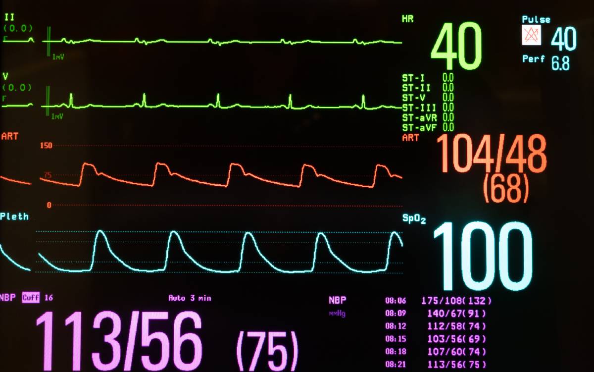
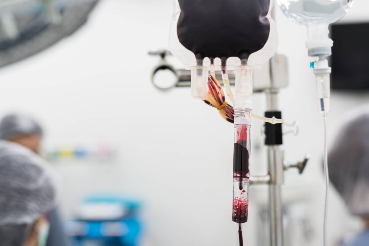
Data on Suzetrigine: A New Non-Opioid Pain Medication

Effective pain management is essential to improving patient comfort, facilitating faster recovery, and reducing the risk of chronic pain development. Multimodal pain management, which involves the use of multiple analgesic agents with different mechanisms of action, has become the standard of care in many clinical settings. Common types of analgesics used in this approach include nonsteroidal anti-inflammatory drugs (NSAIDs), acetaminophen, and opioids. However, the medical field is increasingly seeking non-opioid solutions whenever possible due to opioids’ adverse effects. Recently, research on non-opioid pain medication has seen a major breakthrough with FDA approval of a new drug, suzetrigine, based on its clinical trial data.

Opioids have been a cornerstone of pain management for decades, particularly for moderate to severe acute pain. Their use in medical practice dates back to the early 19th century, when morphine was isolated from opium in 1804. Since then, various opioid medications have been developed, including codeine, hydrocodone, oxycodone, and fentanyl. These drugs have proven highly effective in managing pain but come with significant risks, including addiction, respiratory depression, and other adverse effects.

In response to the ongoing opioid crisis and the need for safer pain management options, researchers have been working to develop novel non-opioid analgesics. One such breakthrough is suzetrigine, marketed under the brand name Journavx. Suzetrigine is a first-in-class, non-opioid pain signal inhibitor that selectively targets the voltage-gated sodium channel NaV1.8. This channel is specifically expressed in peripheral pain-sensing neurons (nociceptors) and plays a crucial role in transmitting pain signals through action potentials. By inhibiting NaV1.8, suzetrigine effectively blocks pain signal transmission without affecting the central nervous system, thereby avoiding the addictive potential and many of the side effects associated with opioids.

On January 30, 2025, the U.S. Food and Drug Administration (FDA) approved suzetrigine (Journavx) for the treatment of moderate to severe acute pain in adult patients. This approval marks a significant milestone in pain management, as suzetrigine represents the first new class of medication for acute pain treatment in over two decades. The FDA’s decision was based on comprehensive data from Phase 2 and 3 clinical trials, which demonstrated the drug’s efficacy and favorable safety profile.

The cost of suzetrigine is significantly higher than traditional opioid medications, with a reported price of $15.50 per pill. This translates to approximately $420 for a one-week course of treatment. Despite the higher cost, suzetrigine may offer long-term savings by reducing the risk of opioid use disorder (OUD) and its associated healthcare costs. Clinical trial data found that the adverse effects of suzetrigine are generally mild and include itching, rash, muscle spasms, and increased levels of creatine kinase. Notably, suzetrigine appears to have a more favorable side effect profile compared to opioids, with less incidence of nausea and drowsiness.

Data have demonstrated the comparative efficacy of suzetrigine against both placebo and opioid analgesics. In two randomized, double-blind, placebo-controlled trials involving patients undergoing abdominoplasty and bunionectomy, suzetrigine showed statistically significant superior pain reduction compared to placebo. When compared to hydrocodone/acetaminophen, suzetrigine demonstrated similar efficacy in pain relief for abdominoplasty patients, although it showed a slower onset of action in bunionectomy patients. These findings suggest that suzetrigine may be a viable alternative to opioids for managing moderate to severe acute pain in multiple surgical settings.

Suzetrigine represents a promising advancement in pain management, offering a non-opioid alternative for patients with moderate to severe acute pain. While its higher cost may present initial challenges for widespread adoption, the potential benefits in terms of reduced opioid-related risks and improved patient outcomes make it a valuable addition to the analgesic armamentarium. As more real-world data becomes available, the role of suzetrigine in clinical practice will likely continue to evolve, potentially reshaping pain management strategies in the years to come.

References

1. U.S. Food and Drug Administration. FDA Approves Novel Non-Opioid Treatment for Moderate to Severe Acute Pain. Published January 30, 2025. Accessed February 5, 2025. doi:10.1001/jama.2025.1234

2. Vertex Pharmaceuticals. Suzetrigine’s Pending Approval Signals a Shift in Non-Opioid Pain Management. Published January 30, 2025. Accessed February 5, 2025. doi:10.1056/NEJMc2025789

3. The New York Times. FDA Approves Journavx Drug to Treat Pain Without Addiction Risk. Published January 30, 2025. Accessed February 5, 2025. doi:10.1136/bmj.k2025.1234

4. Institute for Clinical and Economic Review. Suzetrigine for Acute Pain: Effectiveness and Value. Published December 9, 2024. Accessed February 5, 2025. doi:10.7326/M24-1234

5. HCPLive. FDA Approves Suzetrigine, a Non-Opioid Option, for Treatment of Acute Pain. Published January 30, 2025. Accessed February 5, 2025. doi:10.1001/jamainternmed.2025.5678

Surgery vs. Conservative Treatment for Carpal Tunnel Syndrome

Carpal Tunnel Syndrome (CTS) is a common condition caused by compression of the median nerve as it passes through the carpal tunnel in the wrist. Symptoms often include numbness, tingling, weakness, and pain in the hand and fingers. Choosing the right treatment approach—surgery or conservative measures—depends on the severity of the carpal tunnel syndrome, the patient’s lifestyle, and the desired outcomes.

Conservative treatment is typically the first line of defense, especially for mild to moderate cases, focusing on relieving symptoms and preventing further progression of the condition. Wrist splints, often worn at night, help keep the wrist in a neutral position, reducing pressure on the median nerve and are particularly effective for patients whose symptoms occur primarily during sleep.

Adjusting repetitive hand movements, taking frequent breaks, and improving workplace ergonomics can alleviate symptoms, and avoiding activities that exacerbate symptoms is crucial in this approach. Stretching and strengthening exercises can improve wrist flexibility and reduce inflammation, and therapists may recommend nerve-gliding exercises to ease pressure on the median nerve.

Nonsteroidal anti-inflammatory drugs (NSAIDs) and corticosteroid injections may temporarily reduce inflammation and provide relief, though they are not long-term solutions and work best as part of a broader treatment plan. Acupuncture, yoga, and other holistic approaches have shown promise for some individuals, and while evidence varies, these methods are generally safe and can complement other treatments 1–4.

When conservative treatment fails or symptoms are severe, surgery may be necessary, with the primary goal being to relieve pressure on the median nerve by cutting the transverse carpal ligament, which forms the roof of the carpal tunnel. Open carpal tunnel release, a traditional surgical intervention, involves a small incision in the palm to access and release the ligament, with high success rates. A less invasive option, endoscopic carpal tunnel release, uses smaller incisions and a camera to guide the procedure, offering generally faster recovery times, though with a success rate comparable to open surgery. Post-surgical recovery typically takes a few weeks to a few months, depending on the method and individual factors, and physical therapy is often recommended to regain strength and mobility 5,6.

Conservative treatment methods are best suited for early stage carpal tunnel syndrome or patients who cannot undergo surgery. They are non-invasive, affordable, and have no downtime. However, they may not resolve severe or long-standing cases.

Alternatively, surgery offers a permanent solution for severe CTS but comes with higher costs, potential complications, and longer recovery times. It is usually considered when other treatments fail or when symptoms significantly impair quality of life 7–9.

The decision between surgery and conservative treatment for carpal tunnel syndrome depends on individual circumstances, the severity of symptoms, and the patient’s personal and professional needs. Consulting with a healthcare professional is essential to weigh the benefits and risks of each approach, ensuring the best possible outcome.

References

1.         Surgical versus non-surgical treatment for carpal tunnel syndrome. https://www.cochrane.org/CD001552/NEUROMUSC_surgical-versus-non-surgical-treatment-carpal-tunnel-syndrome DOI: 10.1002/14651858.CD001552.pub3.

2.         Carlson, H. et al. Current options for nonsurgical management of carpal tunnel syndrome. Int J Clin Rheumtol 5, 129–142 (2010). DOI: 10.2217/IJR.09.63

3.         O’Connor, D., Marshall, S. C., Massy‐Westropp, N. & Pitt, V. Non‐surgical treatment (other than steroid injection) for carpal tunnel syndrome. Cochrane Database Syst Rev 2003, CD003219 (2003). DOI: 10.1002/14651858.CD003219

4.         How to Treat Carpal Tunnel Syndrome Without Surgery | The Hand Society. https://www.assh.org/handcare/blog/how-to-treat-carpal-tunnel-syndrome-without-surgery.

5.         Contributors, W. E. Surgery for Treating Carpal Tunnel Syndrome. WebMD https://www.webmd.com/pain-management/carpal-tunnel/do-i-need-carpal-tunnel-surgery.

6.         After Surgery: Discomforts and Complications | Johns Hopkins Medicine. https://www.hopkinsmedicine.org/health/treatment-tests-and-therapies/after-surgery-discomforts-and-complications.

7.         Lusa, V., Karjalainen, T. V., Pääkkönen, M., Rajamäki, T. J. & Jaatinen, K. Surgical versus non-surgical treatment for carpal tunnel syndrome. Cochrane Database Syst Rev 1, CD001552 (2024).

8.         Donati, D., Boccolari, P. & Tedeschi, R. Manual Therapy vs. Surgery: Which Is Best for Carpal Tunnel Syndrome Relief? Life 14, 1286 (2024). DOI: 10.3390/life14101286

9.         Verdugo, R. J., Salinas, R. S., Castillo, J. & Cea, J. G. Surgical versus non-surgical treatment for carpal tunnel syndrome. Cochrane Database Syst Rev CD001552 (2003) DOI: 10.1002/14651858.CD001552.

Anesthesia Research Journals

Research journals are critical for advancing understanding and encouraging innovation in the field of anesthesia. These research journals publish peer-reviewed studies on everything from anesthetic pharmacology and pain management techniques to perioperative patient care and innovations in anesthesia technology. By providing a platform for clinical findings, case studies, and experimental research, academic journals play an essential role in guiding anesthesiologists, researchers, and healthcare professionals toward evidence-based practices and improved patient outcomes.

Several prominent journals lead the field in anesthesia research, each contributing to diverse aspects of anesthesiology. These include Anesthesiology, British Journal of Anaesthesia, Anesthesia & Analgesia, and others.

The journal Anesthesiology, published by the American Society of Anesthesiologists (ASA), is one of the most highly regarded journals in the field. It covers a wide range of topics, including anesthetic techniques, patient safety, critical care, and pain management 1.

The British Journal of Anaesthesia (BJA), one of Europe’s leading journals in anesthesia, publishes research on anesthesia methods, pharmacology, and perioperative medicine. The journal emphasizes both clinical and experimental studies, encouraging high-quality research from an international perspective. Its impact extends into intensive care, reflecting the close relationship between anesthesia and critical care 2.

Anesthesia & Analgesia, supported by the International Anesthesia Research Society, covers a variety of topics, from pharmacology and anesthesia methods to pain management and patient safety. It publishes many high impact articles, including the latest on anesthetic drug development and innovative surgical techniques 3.

The Journal of Clinical Anesthesia provides a platform for practical and clinically relevant research that helps inform everyday anesthetic practice. It aims to provide insights that can directly influence patient care practices in both routine and high-risk cases 4.

Regional Anesthesia and Pain Medicine is a journal with a more specific focus and covers regional anesthesia techniques, analgesia, and chronic pain management. Given the opioid crisis, this journal is particularly valuable for its focus on alternative pain management strategies 5.

Anesthesia research journals significantly impact clinical practices by disseminating new findings that anesthesiologists can implement. For instance, studies published in these journals often provide insights into the effectiveness of new anesthetic drugs, advancements in patient monitoring systems, and improved techniques for managing complex surgical cases. Research on patient safety, such as studies exploring the prevention of postoperative complications or techniques to improve airway management, helps refine best practices and enhance outcomes 6,7.

In recent years, anesthesia research journals have seen increased focus on personalized anesthesia approaches, aiming to tailor anesthetic plans to individual patient profiles. There is also a rising interest in enhanced recovery after surgery (ERAS) protocols, which aim to reduce the postoperative recovery time and improve patient experience. Moreover, journals have

prioritized publishing research on non-opioid alternatives for pain relief, reflecting a global shift toward safer, non-addictive pain management options 8–10.

References

1. Anesthesiology. American Society of Anesthesiologists https://pubs.asahq.org/anesthesiology.

2. Home Page: British Journal of Anaesthesia. https://www.bjanaesthesia.org/.

3. Anesthesia & Analgesia. https://journals.lww.com/anesthesia-analgesia/pages/default.aspx

4. Journal of Clinical Anesthesia. https://www.sciencedirect.com/journal/journal-of-clinical-anesthesia

5. Homepage | Regional Anesthesia & Pain Medicine. Regional Anesthesia & Pain Medicine https://rapm.bmj.com.

6. Chen, L., Li, N. & Zhang, Y. High-impact papers in the field of anesthesiology: a 10-year cross-sectional study. Canadian Journal of Anaesthesia 70, 183 (2022). doi: 10.1007/s12630-022-02363-5

7. Global trends in anesthetic research over the past decade: a bibliometric analysis – Gao – Annals of Translational Medicine. https://atm.amegroups.org/article/view/95637/html. DOI: 10.21037/atm-22-1599

8. Dey, S., Sanders, A. E., Martinez, S., Kopitnik, N. L. & Vrooman, B. M. Alternatives to Opioids for Managing Pain. in StatPearls (StatPearls Publishing, Treasure Island (FL), 2024).

9. Melnyk, M., Casey, R. G., Black, P. & Koupparis, A. J. Enhanced recovery after surgery (ERAS) protocols: Time to change practice? Canadian Urological Association Journal 5, 342 (2011). doi: 10.5489/cuaj.11002

10. Moningi, S., Patki, A., Padhy, N. & Ramachandran, G. Enhanced recovery after surgery: An anesthesiologist’s perspective. Journal of Anaesthesiology, Clinical Pharmacology 35, S5 (2019). DOI: 10.4103/joacp.JOACP_238_16

Prediction of Perioperative Hypotension

The Hypotension Prediction Index (HPI) is an algorithm designed for the prediction of hypotension onset in critically ill or perioperative patients, allowing clinicians to take preventative measures. Hypotension, or low blood pressure, can lead to organ damage, increased morbidity and even mortality, making its timely prediction and management critical. The HPI uses machine learning algorithms to analyze physiological data such as heart rate, blood pressure, and stroke volume to provide clinicians with real-time prediction of hypotensive events before they occur (1). Accurate prediction of perioperative hypotension has significant clinical benefits.

One of the most impactful applications of HPI is in intraoperative care, where patients are particularly vulnerable to blood pressure fluctuations due to anesthesia and other stressors. Prolonged periods of low blood pressure during surgery have been associated with an increased risk of myocardial infarction, stroke, and acute kidney injury (2). For example, Maheshwari et al. demonstrated that hypotension during non-cardiac surgery is independently associated with an increased risk of myocardial injury, suggesting that tight control of blood pressure is essential to minimize these risks (3). HPI addresses this challenge by allowing anesthesiologists to anticipate and prevent hypotension before it reaches critical levels.

A landmark study by Hatib et al. validated the efficacy of the Hypotension Prediction Index in predicting intraoperative hypotension during surgery. The study demonstrated that the HPI can predict hypotension with a sensitivity and specificity greater than 85%, making it a reliable tool in clinical practice (1). In this study, high-fidelity analysis of arterial pressure waveforms was used to train the algorithm, improving the prediction of hypotensive events.

Despite its proven benefits, widespread adoption of HPI in clinical practice has not been without challenges. A key limitation of the system is its reliance on continuous, high-quality hemodynamic monitoring, which is typically only available in well-equipped operating rooms and intensive care units. As a result, not all perioperative settings can use this prediction index to control hypotension. In addition, while the predictive accuracy of HPI is impressive, there is always the potential for false positives. In this case, clinicians may intervene unnecessarily, exposing patients to treatments that may carry their own risks, such as over-administration of fluids or vasopressors (2). Some critics have argued that while HPI is a valuable tool, it should complement, rather than replace, clinical judgment.

In addition, research is ongoing to optimize the use of HPI in different clinical settings. Currently, it is most commonly used in high-risk perioperative and critically ill patients, where real-time blood pressure monitoring is essential and where hypotension prediction is feasible. However, future iterations of the HPI algorithm may incorporate additional physiological parameters or be used in a wider range of medical settings, such as emergency rooms or general wards, where hypotension also poses a significant risk (3).

In conclusion, the Hypotension Prediction Index represents a leap forward in the management of hypotension in perioperative and critical care settings. By using machine learning algorithms

to predict hypotension before it occurs, the HPI enables timely interventions that can prevent adverse outcomes. While challenges related to data quality and clinical interpretation remain, the growing body of evidence supporting the efficacy of HPI suggests that it will play an increasingly important role in improving patient outcomes and reducing the risks associated with hypotension.

References

1. Hatib F, Jian Z, Buddi S, et al. Machine-learning Algorithm to Predict Hypotension Based on High-fidelity Arterial Pressure Waveform Analysis. Anesthesiology. 2018;129(4):663-674. doi:10.1097/ALN.0000000000002300

2. Davies SJ, Vistisen ST, Jian Z, Hatib F, Scheeren TWL. Ability of an Arterial Waveform Analysis-Derived Hypotension Prediction Index to Predict Future Hypotensive Events in Surgical Patients. Anesth Analg. 2020;130(2):352-359. doi:10.1213/ANE.0000000000004121

3. Maheshwari K, Turan A, Mao G, et al. The association of hypotension during non-cardiac surgery, before and after skin incision, with postoperative acute kidney injury: a retrospective cohort analysis. Anaesthesia. 2018;73(10):1223-1228. doi:10.1111/anae.14416

Anesthesia Considerations for Aspirin

Aspirin is widely used for its antiplatelet effects in the prevention of cardiovascular events, but it also raises specific considerations in the context of anesthesia due to its impact on bleeding risk. The primary concern with aspirin in the perioperative setting is its irreversible inhibition of cyclooxygenase-1 (COX-1), which leads to decreased thromboxane A2 production, impairing platelet aggregation and prolonging bleeding time (1). This effect requires careful management strategies, particularly when neuraxial anesthesia or other regional anesthetic techniques are planned, as these are associated with a risk of hematoma formation.

The American Society of Regional Anesthesia and Pain Medicine (ASRA) guidelines indicate that low-dose aspirin therapy does not significantly increase the risk of spinal hematoma following epidural or spinal anesthesia and can generally be continued in the perioperative period (2). However, other studies emphasize the need for vigilance and individualized assessment, especially in patients receiving higher doses or those with other risk factors for bleeding (3). The decision to continue or discontinue aspirin therapy must balance the risk of thrombosis against the potential for bleeding complications, taking into account the specific surgical procedure, patient comorbidities, and type of anesthesia.

The perioperative continuation of aspirin in patients at risk for vascular complications, such as those with coronary stents, is supported by evidence that the benefits of maintaining aspirin therapy generally outweigh the risks (4). Discontinuation of aspirin therapy can lead to a rebound prothrombotic state, increasing the likelihood of myocardial infarction or stroke, particularly in high-risk patients (5). This is particularly relevant in non-cardiac surgery, where the risk of bleeding is lower, and the consequences of a thrombotic event can be severe.

On the other hand, for procedures where major bleeding would pose significant risks, such as intracranial procedures, the decision-making process becomes more complex. In these scenarios, some guidelines suggest stopping aspirin at least seven days before surgery and anesthesia to allow for sufficient platelet recovery, while others emphasize a case-by-case approach (1). This is further complicated by the lack of a reliable, rapid method to reverse aspirin’s effects, unlike other anticoagulants for which specific reversal agents exist.

The importance of interdisciplinary communication cannot be overstated in managing anesthesia for patients on aspirin therapy. Anesthesiologists must work closely with surgeons and the patient’s primary care or cardiology teams to evaluate the risks and benefits of continuing aspirin therapy perioperatively. The anesthetic plan should include strategies for managing potential bleeding, such as availability of blood products and the use of less invasive surgical techniques where feasible.

Anesthesia considerations for patients on aspirin therapy require a nuanced approach that weighs the thrombotic risks of discontinuation against the bleeding risks of continuation. Guidelines generally support the continuation of low-dose aspirin for most surgeries, especially where the risk of major bleeding is low, but emphasize individual risk assessment for each patient. Anesthesia providers must remain vigilant and adaptable, employing a multidisciplinary approach to optimize patient outcomes.

References

  1. Song JW, Soh S, Shim JK. Dual antiplatelet therapy and non-cardiac surgery: evolving issues and anesthetic implications. Korean J Anesthesiol. 2017.
  2. Horlocker TT, Wedel DJ, Benzon H, et al. Regional anesthesia in the anticoagulated patient: defining the risks (the second ASRA Consensus Conference on Neuraxial Anesthesia and Anticoagulation). Reg Anesth Pain Med. 2003.
  3. Macdonald R. Aspirin and extradural blocks. Br J Anaesth. 1991;66(1):1-6.
  4. Vela Vásquez RS, et al. Aspirin and spinal haematoma after neuraxial anaesthesia: Myth or reality? Br J Anaesth. 2015;115(5):688-695.
  5. Devereaux PJ, Mrkobrada M, Sessler DI, et al. Aspirin in patients undergoing noncardiac surgery. N Engl J Med. 2014;370(16):1494-1503

The Effect of Propofol on Neurotransmitters

Loss of consciousness (LOC) is a medical event characterized by a disruption in someone’s awareness and responsiveness to their environment. Anesthesia providers often intentionally induce LOC during surgical procedures using anesthetics such as propofol. Decades of research have aimed to shed light on the neural mechanisms behind anesthesia-induced LOC, including the associated neuroanatomy and neurotransmission. Studies suggest that propofol, a widely used anesthetic, exerts its effects via the neurotransmitters dopamine, GABA, and glutamate.

Propofol is a well-established intravenous anesthetic known for its smooth induction, rapid terminal half-life, and low incidence of postoperative nausea and vomiting.1 Dynamic causal modeling of neural activity suggested that propofol-induced LOC impaired backward connectivity from frontal to parietal cortices. Additional Bayesian model selection alluded that backward connectivity in higher-order associative cortical regions may be a crucial bottleneck for conscious awareness.2 Through an in-depth analysis into these higher-order associative areas, studies found the prefrontal cortex (PFC), and especially the medial prefrontal cortex, displayed significant changes in neurotransmission during anesthesia induction and emergence. As such, it is suggested that propofol induces LOC at least partially through impacts on the PFC.3

Studies on nicotine abuse have demonstrated the impact of dopamine firing in the ventral tegmental area (VTA) on the modulation of extracellular dopamine levels in the PFC. In a 2016 study, researchers performed in vivo and in vitro analysis on 90 rats, including observation, microdialysis, and histology. Intravenous administration of propofol and the subsequent LOC caused a significant increase in the PFC levels of the dopamine metabolite 3,4-Dihydroxyphenylacetic acid (DOPAC), while decreasing the PFC levels of dopamine neurotransmitters (NT). Both DOPAC and dopamine NT levels returned to baseline after emergence from anesthesia. Therefore, it is proposed propofol induces LOC through facilitating the catabolism of dopamine NT into its metabolite, DOPAC, in the PFC.4 

Since propofol is known to exert its anesthetic effects through the inhibitory neurotransmitter GABA, the above researchers blocked the GABAA receptors in the PFC and found delayed induction of LOC and hastened emergence from LOC. Since blocking GABA reduces (but does not eliminate) the effect of propofol, these results indicate propofol induces LOC at least partially through the GABA neurotransmitters and GABAA receptors in the PFC.

In early 2009, 10 healthy human volunteers (aged 20- 40) participated in a noninvasive magnetic resonance spectroscopy study. At a propofol injection concentration of 1.5 μg/ml, subjects began showing sedative qualities; by 3.0 μg/ml, they lost consciousness. During LOC, the researchers observed significantly decreased levels of glutamate, the brain’s primary excitatory neurotransmitter, in all patients, along with up-regulated levels of GABA.5

The effects of propofol on neurotransmitters during loss of consciousness highlight the drug’s profound effect on neural activity. By modulating critical neurotransmitters like dopamine, GABA, and glutamate in the PFC, propofol can induce LOC, which is essential for various medical procedures. Understanding these pharmacological mechanisms not only enhances our ability to use propofol safely and effectively, but also provides invaluable insights into the neural processes underlying consciousness. Continued research in this area is key to advancing anesthetic practices and improving patient outcomes. 

References 

  1. Sahinovic, Marko M., et al. “Clinical Pharmacokinetics and Pharmacodynamics of Propofol.” Clinical Pharmacokinetics, vol. 57, no. 12, Dec. 2018, pp. 1539–58. https://doi.org/10.1007/s40262-018-0672-3
  2. Boly, Mélanie, et al. “Connectivity Changes Underlying Spectral EEG Changes During Propofol-Induced Loss of Consciousness.” Journal of Neuroscience, vol. 32, no. 20, May 2012, pp. 7082–90. https://doi.org/10.1523/JNEUROSCI.3769-11.2012
  3. Leon-Dominguez, Umberto, et al. “Molecular Concentration of DeoxyHb in Human Prefrontal Cortex Predicts the Emergence and Suppression of Consciousness.” NeuroImage, vol. 85, Jan. 2014, pp. 616–25. https://doi.org/10.1016/j.neuroimage.2013.07.023
  4. Wang, Yuan, et al. “Effects of Propofol on the Dopamine, Metabolites and GABAA Receptors in Media Prefrontal Cortex in Freely Moving Rats.” American Journal of Translational Research, vol. 8, no. 5, May 2016, pp. 2301–08. https://www.ncbi.nlm.nih.gov/pmc/articles/PMC4891442/
  5. Zhang, Hui, et al. “Effect of Propofol on the Levels of Neurotransmitters in Normal Human Brain: A Magnetic Resonance Spectroscopy Study.” Neuroscience Letters, vol. 467, no. 3, Dec. 2009, pp. 247–51. https://doi.org/10.1016/j.neulet.2009.10.052

Pitting Edema: Considerations for the OR

Edema is characterized by a noticeable swelling caused by the expansion of interstitial fluid volume. Pitting edema is identified by a depression in the tissue that remains for at least five seconds after applying pressure to the swollen area. This type of edema indicates the movement of excess interstitial water in response to pressure. A number of conditions affecting the circulatory and/or lymphatic system can cause pitting edema, most often in peripheral areas. Signs of pitting edema indicate potential risk factors for surgery and anesthesia and the need for the OR team to investigate the patient’s condition and medical history.

Peripheral edema typically occurs in dependent areas, appearing mainly in the lower extremities of ambulatory patients and over the sacrum in bedridden patients. It can also occur in the upper extremities, though less frequently. Several clinical conditions are linked to the development of edema, including heart failure, cirrhosis, nephrotic syndrome, and other conditions such as venous and lymphatic diseases. Determining the cause of edema depends on whether it is unilateral or bilateral. The sudden onset of unilateral leg edema often raises concerns about deep vein thrombosis (DVT). In addition to edema, DVT patients may experience calf tenderness, pain, or firmness along a vein, or unilateral warmth or erythema. The most common cause of chronic unilateral or asymmetric edema is chronic venous disease in the lower extremities. Acute bilateral leg edema is rare and may be due to medications, acute heart failure, or acute nephrotic syndrome. Chronic bilateral leg edema is usually caused by chronic venous disease, although heart failure and pulmonary hypertension are often underdiagnosed. Less common causes of chronic bilateral leg edema include renal and liver diseases.

If present, pitting edema should be assessed by the OR team before surgery due to its association with increased perioperative complications. For instance, patients with pitting edema caused by a DVT need anticoagulation therapy to lower the risk of recurrent DVT, heart attack, and stroke. However, while on blood thinners, these patients face an increased risk of surgical bleeding. Therefore, it is advisable to delay surgery if the patient is clinically stable or to consider withholding anticoagulation during the perioperative period.

In patients with bilateral edema due to liver disease, evaluating surgical risk involves assessing the severity of liver disease, the urgency of surgery (and alternatives to surgery), and any coexisting medical conditions. Surgery is contraindicated in patients with acute liver failure, alcoholic hepatitis, and severe chronic hepatitis, as the risks surpass the benefits. For patients with less severe liver disease, physicians commonly use surgical risk calculators, such as the Child-Turcotte-Pugh score, to determine if the benefits of surgery outweigh the risks.

Finally, for patients with pitting edema due to heart failure, obtaining a chest X-ray can help determine the presence of pulmonary edema (fluid accumulation in the lungs), which significantly increases surgical risk. Patients experiencing an acute exacerbation of heart failure may have shortness of breath due to pulmonary edema, and this condition should be managed with diuretics before surgery to prevent cardiopulmonary complications.

Lastly, the OR and PACU teams should recognize that pitting edema may worsen after surgery due to the release of inflammatory signals that increase capillary permeability, allowing more fluid to accumulate in the interstitial space. Additionally, patients often receive IV fluids during the perioperative period, which can further exacerbate edema. Ultimately, the decision to proceed with surgery is influenced by the urgency of the procedure (emergency, urgent, or elective), the severity of symptoms, the presence of comorbid conditions, and the specific risks associated with the proposed surgery.

References

Lawenda BD, Mondry TE, Johnstone PA. Lymphedema: a primer on the identification and management of a chronic condition in oncologic treatment. CA Cancer J Clin. 2009 Jan-Feb;59(1):8-24. doi: 10.3322/caac.20001. PMID: 19147865.

Hull R, Hirsh J, Sackett DL, Taylor DW, Carter C, Turpie AG, Powers P, Gent M. Clinical validity of a negative venogram in patients with clinically suspected venous thrombosis. Circulation. 1981 Sep;64(3):622-5. doi: 10.1161/01.cir.64.3.622. PMID: 7261292.

Gorman WP, Davis KR, Donnelly R. ABC of arterial and venous disease. Swollen lower limb-1: general assessment and deep vein thrombosis. BMJ. 2000 May 27;320(7247):1453-6. doi: 10.1136/bmj.320.7247.1453. PMCID: PMCID: PMC1127644.

Blankfield RP, Finkelhor RS, Alexander JJ, Flocke SA, Maiocco J, Goodwin M, Zyzanski SJ. Etiology and diagnosis of bilateral leg edema in primary care. Am J Med. 1998 Sep;105(3):192-7. doi: 10.1016/s0002-9343(98)00235-6. PMID: 9753021.

Guazzi M, Polese A, Magrini F, Fiorentini C, Olivari MT. Negative influences of ascites on the cardiac function of cirrhotic patients. Am J Med. 1975 Aug;59(2):165-70. doi: 10.1016/0002-9343(75)90350-2. PMID: 1155476.

Matthay MA. Resolution of pulmonary edema. Thirty years of progress. Am J Respir Crit Care Med. 2014 Jun 1;189(11):1301-8. doi: 10.1164/rccm.201403-0535OE. PMID: 24881936; PMCID: PMC4098087.

Hypocalcemia and Anesthesia

Calcium is crucial for various physiological processes, especially muscle contraction. In skeletal muscle, calcium release from the sarcoplasmic reticulum initiates an action potential leading to muscle contraction. Calcium also triggers contractions in smooth and cardiac muscle. Intercalated discs enable the transmission of contraction signals between cardiac myocytes, leading to synchronized muscle contractions throughout the heart. This coordination ensures that electrical impulses travel from the atria to the ventricles, driving blood flow through the circulatory system. Disruptions in calcium levels, such as hypocalcemia, can interfere with these normal physiological processes and impact anesthesia protocols.

Acute hypocalcemia (below 8.5 mg/dL) can cause syncope, congestive heart failure, numbness and tingling, bronchospasm and wheezing, laryngospasm and dysphagia, irritability, depression, fatigue, and seizures. The hallmark of acute hypocalcemia is tetany, characterized by neuromuscular irritability. Electromyographically, tetany involves repetitive, high-frequency discharges following a single stimulus. Peripheral neuron hyperexcitability is a significant pathophysiological effect of hypocalcemia, occurring at all levels of the nervous system, including motor endplates, spinal reflexes, and the central nervous system. Chronic hypocalcemia can result in coarse hair, brittle nails, psoriasis, dry skin, pruritus, poor dentition, and cataracts. Common physical exam findings of hypocalcemia include neural hyperexcitability, psychological disturbances, and cardiac arrhythmias.

The major factors influencing serum calcium concentration are parathyroid hormone (PTH), vitamin D, fibroblast growth factor 23 (FGF23), the calcium ion itself, and phosphate. Low serum calcium levels are most often caused by disorders of PTH or vitamin D. Hypocalcemia with low PTH occurs when there is decreased secretion of PTH due to destruction of the parathyroid glands (e.g. postsurgical, autoimmune), abnormal parathyroid gland development, or altered regulation of PTH production and secretion. Among the various causes of hypocalcemia, postsurgical hypoparathyroidism and autoimmune hypoparathyroidism are two of the more common ones. Hypocalcemia with high PTH occurs when PTH rises in response to low serum calcium levels to mobilize calcium from the kidneys and bones and to increase vitamin D production. Chronic hypocalcemia occurs when these actions are inadequate to restore serum calcium to normal levels. A high serum PTH in a patient with hypocalcemia may be secondary to vitamin D deficiency, chronic kidney disease, or pseudohypoparathyroidism, which is a rare condition.

During anesthesia, various factors can alter serum ionized calcium levels, thereby increasing the risk of adverse effects from hypocalcemia in susceptible patients. These factors include malnutrition and low albumin levels, abnormal acid-base balance and electrolyte levels, medications used during the peri-operative period, transfusion of large volumes of citrated blood, and the use of cardiopulmonary bypass. Anesthetists should aim to prevent further changes in plasma calcium concentration and promptly recognize and treat the adverse effects of hypocalcemia, especially those affecting the heart. Preoperative evaluations typically include an assessment of electrolytes, including calcium, though abnormalities may potentially also arise intraoperatively or postoperatively.

The treatment of hypocalcemia depends on its severity and underlying cause. Patients with severe symptoms (such as bronchospasm, seizures, or decreased cardiac function) require rapid correction with intravenous calcium therapy. For those with milder symptoms of neuromuscular irritability, initial treatment with oral calcium supplementation is sufficient. Intravenous calcium is also indicated to prevent acute hypocalcemia in patients with mild hypocalcemia or chronic hypocalcemia who cannot take or absorb oral supplements, often due to complex surgical procedures requiring prolonged recovery.

In conclusion, understanding the role of calcium in physiological processes, the diverse factors influencing its concentration, and the appropriate management strategies for hypocalcemia is essential for ensuring optimal patient care, particularly for anesthesia and surgery.

References

Riccardi D, Brown EM. Physiology and pathophysiology of the calcium-sensing receptor in the kidney. Am J Physiol Renal Physiol. 2010 Mar;298(3):F485-99. doi: 10.1152/ajprenal.00608.2009. PMID: 19923405; PMCID: PMC2838589.

Hannan FM, Thakker RV. Investigating hypocalcaemia. BMJ. 2013 May 9;346:f2213. doi: 10.1136/bmj.f2213. PMID: 23661111.

Desai TK, Carlson RW, Geheb MA. Prevalence and clinical implications of hypocalcemia in acutely ill patients in a medical intensive care setting. Am J Med. 1988 Feb;84(2):209-14. doi: 10.1016/0002-9343(88)90415-9. PMID: 3407650.

Zivin JR, Gooley T, Zager RA, Ryan MJ. Hypocalcemia: a pervasive metabolic abnormality in the critically ill. Am J Kidney Dis. 2001 Apr;37(4):689-98. doi: 10.1016/s0272-6386(01)80116-5. PMID: 11273867.

Kluger MT, Tham EJ, Coleman NA, Runciman WB, Bullock MF. Inadequate pre-operative evaluation and preparation: a review of 197 reports from the Australian incident monitoring study. Anaesthesia. 2000 Dec;55(12):1173-8. doi: 10.1046/j.1365-2044.2000.01725.x. PMID: 11121926.

Blitz JD, Kendale SM, Jain SK, Cuff GE, Kim JT, Rosenberg AD. Preoperative Evaluation Clinic Visit Is Associated with Decreased Risk of In-hospital Postoperative Mortality. Anesthesiology. 2016 Aug;125(2):280-94. doi: 10.1097/ALN.0000000000001193. PMID: 27433746.

Training Requirements for CRNAs

Certified Registered Nurse Anesthetists (CRNAs) are advanced practice nurses who specialize in providing anesthesia care to patients across a wide range of clinical settings. Their role is crucial in ensuring safe and effective anesthesia administration during surgical procedures, obstetric deliveries, and pain management interventions. CRNAs must fulfill rigorous and comprehensive training requirements that encompass both academic education and clinical experience 1.

The path to becoming a CRNA typically begins with earning a Bachelor of Science in Nursing (BSN) degree from an accredited nursing program. This undergraduate education provides aspiring CRNAs with a strong foundation in nursing theory, patient care principles, and basic sciences. During their BSN program, students may have the opportunity to gain exposure to anesthesia-related topics through elective courses or clinical rotations 2.

Following completion of their BSN degree, aspiring CRNAs must obtain a valid Registered Nurse (RN) license by passing the National Council Licensure Examination for Registered Nurses (NCLEX-RN). This licensure is a prerequisite for entering graduate-level nurse anesthesia programs, which are accredited by the Council on Accreditation of Nurse Anesthesia Educational Programs (COA) 2.

The core component of training requirements for CRNAs is a graduate-level education program in nurse anesthesia that awards a nurse anesthesia practice (DNAP) or a Doctor of Nursing Practice (DNP) degree. Depending on the institution and the level of the degree, these programs typically require full-time enrollment and last a few years 2.

CRNA educational programs are intensive and interdisciplinary, covering a wide range of topics essential for anesthesia practice. Coursework may include advanced physiology and pharmacology, principles of anesthesia practice, anesthesia equipment and technology, patient assessment and monitoring, anesthesia techniques and procedures, and professional ethics and standards. Students also receive instruction in advanced nursing concepts, leadership skills, and evidence-based practice 3.

In addition to classroom instruction, training requirements for CRNAs include extensive clinical practicum experience under the supervision of qualified preceptors. These clinical rotations take place in diverse healthcare settings, including hospitals, ambulatory surgical centers, obstetric units, and pain management clinics. Through hands-on experience, students develop proficiency in anesthesia delivery, airway management, patient monitoring, and crisis management, preparing them for independent practice as CRNAs 3.

Upon successful completion of their graduate program, CRNA graduates must pass the National Certification Examination (NCE), administered by the National Board of Certification and Recertification for Nurse Anesthetists (NBCRNA). This rigorous exam assesses the candidate’s knowledge, skills, and competencies in anesthesia practice and is a prerequisite for obtaining certification as a CRNA 4.

Once certified, CRNAs must maintain their credentials through continuing education and professional development activities. This includes participation in continuing education courses, attendance at conferences and workshops, and engagement in quality improvement initiatives within their practice settings 5.

The training requirements for CRNAs are extensive and demanding, encompassing academic education, clinical experience, and national certification. Through a combination of rigorous coursework and hands-on clinical training, aspiring CRNAs acquire the knowledge, skills, and competencies necessary for safe and effective anesthesia practice. By adhering to high standards of education and professional development, CRNAs uphold the highest standards of patient care and contribute to the delivery of quality anesthesia services across healthcare settings.

References

1.        AANA | How to Become a CRNA. Available at: https://www.aana.com/about-us/about-crnas/become-a-crna/. (Accessed: 28th April 2024)

2.        How To Become A Nurse Anesthetist | NurseJournal.org. Available at: https://nursejournal.org/nurse-anesthetist/how-to-become-a-crna/. (Accessed: 28th April 2024)

3.        How To Become a Nurse Anesthetist (CRNA) in 6 Steps | Indeed.com. Available at: https://www.indeed.com/career-advice/career-development/how-to-become-a-crna. (Accessed: 28th April 2024)

4.        Requirements to Practice as a Nurse Anesthetist in the United States – Council on Accreditation. Available at: https://www.coacrna.org/about-coa/requirements-to-practice-as-a-nurse-anesthetist-in-the-united-states/. (Accessed: 28th April 2024)

5.        AANA | Continuing Education. Available at: https://www.aana.com/ce/. (Accessed: 28th April 2024)

Neuraxial Anesthesia in Patients with Scoliosis

Scoliosis is an abnormal curvature of the spine in the coronal, axial, and/or sagittal planes. The condition is most frequently idiopathic and diagnosed in adolescence, but it can also be a congenital or neuromuscular condition. According to the National Scoliosis Foundation, scoliosis patients make up over 600,000 of private physician office visits, 30,000 children are fitted with a brace, and 38,000 patients undergo spinal fusion surgery per year.1 Naturally, this deformation of the spine can have an effect on the administration and spread of neuraxial, or spinal, anesthesia, with disparities in spread being a well-recognized complication for patients with scoliosis.

As reported by a pilot study by Ballarapu et al. in 2020 in the Indian Journal of Anaesthesia, many anesthesiologists are reluctant to administer neuraxial anesthesia in patients with scoliosis out of fear of postoperative complications (including potential neurological deficits), needing multiple attempts, and the unpredictability in the level and pattern of blockade during spinal anesthesia in these patients.2 Given the prevalence of scoliosis in our population and these known potential complications, it is important for surgeons and anesthesiologists to be familiar with the differences of neuraxial anesthesia in patients with scoliosis, as well as to work to develop a more sophisticated standard of care for these patients. 

A 2013 paper in Regional Anesthesia by researchers from the Vanderbilt University School of Medicine reports on a case in which computerized tomography was used to assist in the placement of an epidural catheter in a patient with severe scoliosis and congenital dwarfism. An anesthesiologist typically administers neuraxial anesthesia by using spinal landmarks as guidance or by using ultrasound guidance. Due to the risks posed in trying to administer neuraxial anesthesia to a patient with such pronounced scoliosis (including neural injury, spinal hematoma, post-dural puncture headache, or infection as well as, more generally, a decrease in procedure efficiency and increase in patient discomfort and decrease in patient satisfaction), the computerized tomography data, corroborated by fluoroscopic images and ultrasound, were determined to be necessary precautions prior to operating on this patient. The surgical procedure was conducted without complications, and there were no adverse effects of the surgery. The authors suggested an algorithm to guide neuraxial techniques in scoliotic patients, taking the etiology and severity of a patient’s scoliosis into account in determining what techniques may be necessary in reducing risk of injury or discomfort for the patient in administering neuraxial anesthesia.3

A later (2016) report by Sharma and McConachie in Journal of Obstetric Anaesthesia and Critical Care reviewed further findings and techniques on how to safely administer neuraxial anesthesia in scoliotic patients. Noting that neuraxial blocks, despite being the preferred form of anesthesia for parturient patients in particular, were historically avoided in patients who had scoliosis or previous back surgery due to its comparative difficulty, higher chance of complications, and decreased efficacy, these authors also spotlight the use of ultrasound technology as a potential key element in overcoming these difficulties in providing neuraxial blocks to patients with scoliosis. They found through their review of recent literature that the use of ultrasound reduced the number of attempts and necessary levels of epidural catheter placement for all parturient patients, including those with a history of severe scoliosis or scoliosis repair. By allowing for the identification of interspinous spaces, ultrasound assisted with the insertion angle of the Tuohy needle and with the identification of the epidural space in these cases.4

All in all, as detailed in a 2020 review on the topic,5 ultrasound-guided placement of neuraxial blocks has become common practice for patients with severe scoliosis. Using this technique, it is possible to minimize the risk of misplacement, complications, or postoperative pain. Due to this technique becoming commonplace, and due to the prevalence of patients with either scoliosis or other conditions that increase the difficulty of administering neuraxial anesthesia (such as obesity, prior history of back surgery, or non-scoliotic abnormalities of the spine), it is important that anesthesiologists who routinely perform lumbar neuraxial blocks be familiar with both the sonoanatomy of the lumbar vertebrae and the techniques for ultrasound-guided placement of neuraxial blocks.

References

(1)  Scoliosis – Symptoms, Diagnosis and Treatment. https://www.aans.org/en/Patients/Neurosurgical-Conditions-and-Treatments/Scoliosis.

(2)  Ballarapu, G.; Nallam, S.; Samantaray, A.; Kumar, V. K.; Reddy, A. Thoracolumbar Curve and Cobb Angle in Determining Spread of Spinal Anesthesia in Scoliosis. An Observational Prospective Pilot Study. Indian J Anaesth 2020, 64 (7), 594. https://doi.org/10.4103/ija.IJA_914_19.

(3)  Bowens, C.; Dobie, K. H.; Devin, C. J.; Corey, J. M. An Approach to Neuraxial Anaesthesia for the Severely Scoliotic Spine. Br J Anaesth 2013, 111 (5), 807–811. https://doi.org/10.1093/bja/aet161.

(4)  Sharma, M.; McConachie, I. Neuraxial Blocks in Parturients with Scoliosis and after Spinal Surgery. J Obstet Anaesth Crit Care 2016, 6 (2), 70. https://doi.org/10.4103/2249-4472.191594.

(5)  Yoo, S.; Kim, Y.; Park, S.-K.; Ji, S.-H.; Kim, J.-T. Ultrasonography for Lumbar Neuraxial Block. Anesth Pain Med 2020, 15 (4), 397–408. https://doi.org/10.17085/apm.20065.